The visual system transmits more than 90% of sensory information to the brain. Vision is a multi-unit process, starting from the projection of the image on the retina, then the photoreceptors are excited, the transmission and transformation of visual information in the neural layers of the visual system. The visual perception ends with the formation of the large hemispheres of the visual image in the occipital lobe of the cortex.
The cones are mainly located in the recess of the retina, called fovea, which is the point of greatest visual acuity in which there are about seven million cones. The rays of light are reflected in the foveas, reflected by the object that we are looking at.
In the first case, the light that comes from the objects in question is refracted by the cornea, enters through the pupil and is again refracted by the crystalline lens. The rays of light finally converge in the retina, intersecting the various layers that are in it, until they reach the photoreceptor cells.
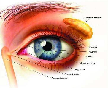
The peripheral part of the visual analyzer is represented by the organ of sight (the eye), which serves to perceive light stimuli and is located in the orbit. The organ of vision consists of an eyeball and an auxiliary device (Diagram 12.1). The structure and function of the organ of vision are presented in table 12.1.
Scheme 12.1.
The structure of the organ of vision
The structure of the organ of vision
Auxiliary apparatus
Then, the light energy activates the photopigments, which are located in the photoreceptor membranes, which close the sodium ion channels, which usually open in dark conditions. Therefore, the negativity of the receptor cell or hyperpolarization increases due to a decrease in the consumption of sodium ions, creating photoreceptors of receptor potential, which subsequently becomes a nerve impulse in bipolar neurons.
The optical nerve, a structure that divides into two optical fibers, transmits nerve impulses. For this, it includes half with axons of the nasal half and the other with that of the temporary half of the retina. The axons of the temporary half reach the nuclei of the thalamus on one side, and those of the nasal halves intersect in the optical chiasm, reaching the opposite side of the nuclei of the thalamus.
Eyeball
eyelids with eyelashes
tear glands
outer (protein) shell,
medium (vascular) membrane
inner (retina) shell
Table 12.1.
The structure and function of the eye
|
Systems |
Parts of the eye Finally, the nerve pathways carry nerve impulses into the visual cortex, located in the occipital lobe of each hemisphere of the brain, from the thalamus. A normal eye is called emmetropic. In a normal eye, light rays are focused directly on the retina, however, when there are changes in the diameter of the eyeball or problems in any of its constituent structures, some pathology may occur. Myopia is a condition of the eye, which is caused by the fact that light cannot focus on the retina, which makes it impossible to clearly see distant objects. This is due to the fact that the eye is longer or because the lens is thicker than usual, which increases its converging force. This causes the image to form in front of the retina, so people suffering from this pathology must be very close to the objects so that the image coincides in the retina. |
Structure |
Functions |
|
Auxiliary |
Hair growing from the inner to the outer corner of the eye on the eyebrow |
Take away the sweat from his forehead |
|
|
Skin folds with eyelashes |
Protect the eye from wind, dust, bright sunshine Hyperopia is a pathology of the eye, in which the light is not properly refracted, therefore the images are not clearly focused, being behind the retina. This usually happens when the eye is shorter. Therefore, people with this disease should squint or move away so that the image matches the retina, as they do not see dense objects well. Vision in low light conditions occurs in humans. When the anteroposterior axis of the eye is extended, an image is formed in front of the retina. This abnormality of vision is known as. The retina is the layer that internally covers the eye chamber and consists of two types of cells, cones and rods. According to your knowledge of cones, check out the wrong alternative. |
||
|
Lacrimal apparatus |
Lacrimal glands and tear paths |
Tears moisturize the surface of the eye, clean, disinfect (lysozyme) and warm it |
|
|
Shell |
Protein |
Outer dense shell consisting of connective tissue Look at the following images and check the right alternative. Observe the anatomy of the eye and check the correct alternative. The retina is the layer that internally covers the eye chamber and contains two types of cells that are stimulated by light, rods and cones. The human eye is covered with a sclera, a protective layer of fibrous connective tissue that is transparent in front of the eye, where it forms the cornea. The diaphragm is located in the front of the choroid and is responsible for the color of the eyes, avoiding reflections of light that prevent the formation of a clear image. |
Eye protection against mechanical and chemical damage, as well as microorganisms |
|
Vascular |
Median sheath, riddled with blood vessels. The inner surface of the shell contains a layer of black pigment |
The power of the eye, the pigment absorbs light rays |
|
|
Retina The viewer is in the center of the iris, and this is the hole through which the light passes. The lens is a protein structure in the form of a biconvex lens, which gives sharpness and focus on the luminous image formed in the cornea. Currently, the lens is called by many authors of lenses. The rods are extremely photosensitive photodetectors, but cannot distinguish colors. The cones are less sensitive to light than the rods, but they have the ability to distinguish different wavelengths, providing color vision. In dimly lit environments, only more sensitive rods are stimulated. That is why in the penumbra we cannot distinguish the colors of objects, but as the brightness increases, the cones are activated and the colors become visible. |
The inner multilayer shell of the eye, consisting of photoreceptors: rods and cones. There is a blind spot in the back of the retina (there are no photoreceptors) and a yellow spot (the highest concentration of photoreceptors) |
The perception of light, its transformation into nerve impulses |
|
|
Optical Presbyopia: Also called visual fatigue, occurs as you age. This is caused by a loss of capacity in the crystalline arrangement; it can be corrected with converging lenses. Hyperopia: the eyeball is shorter than usual, so images of neighboring objects are formed after the retina. This problem can be corrected with the use of converging lenses. Myopia: The eyeball is more elongated than usual, which prevents proper targeting of more distant objects. In myopia, the image is focused in front of the retina. Correction is made using a diverging lens. Astigmatism: astigmatism is due to asymmetry of corneal curvature or, more rarely, lens curvature. This causes some images to be projected without sharpness on the retina. Correction of this problem is carried out with the help of cylindrical lenses that have uneven curvatures, compensating for the uneven curvature of the eye. |
Cornea |
Transparent front part of the tunica |
Refrains light rays |
|
Watery moisture |
Clear fluid behind the cornea |
Transmits the rays of light |
|
|
Anterior choroid with pigment and muscle Communication problems arise from the first year of age, when they seriously affect the development of a child in all areas. It is very important to assess the visual abilities of these children as soon as possible, and exams should be repeated at regular intervals to guide caregivers, therapists and parents, and residual visual capabilities should be used optimally. other ways of communication and education. We often believe that these children are badly damaged by the brain, but some of them have normal brain development, despite serious sensory impairments. Communication works in both directions: most of the disabled are often adults. |
The pigment gives color to the eye (in the absence of pigment the eyes are red in albino), the muscles change the size of the pupil |
||
|
The hole in the center of the iris |
Expanding and tapering, regulates the amount of incoming light in the eye. |
||
|
Lens To understand these malformations, a brief description of the development of the eyeball is needed. At the beginning of its embryonic development, the eyeball looks like a finger-like structure on the surface of the neural tube in an embryo 4 mm long. At the end of this continuation, a depression is formed, then it becomes asymmetrical and invaginates on the side, which will later be the lower part of the eye. These vessels introduce this invagination into the cup-shaped structure, which forms the vessels, do not enter the retina itself, but into the tissue that fills the font, and then disappear when the vitreous gel forms in the posterior chamber, and the remains of these vessels are sometimes visible on the disk. optical in the normal eye. |
Biconvex elastic transparent lens surrounded by ciliary muscle (formation of choroid) |
Refracts and focuses the rays. It has accommodation (the ability to change the curvature of the lens) |
|
|
Vitreous body |
Clear gelatinous substance |
Fills the eyeball. Supports intraocular pressure. Transmits the rays of light As a rule, invagination, through which the vascular pedicle closes in the eye, is closed. Sometimes it does not close either at the level of the front camera, or at the level of the rear camera, or at both. The lack of closure in the front of the eye is responsible for the absence of a substance in the lower part of the retina and choroid, called a coloboma, which is often associated with the absence of a substance in the lower part of the iris, causing the pupil to form a "keyhole". If the lack of closure affects the back of the eye, the coloboma touches the retina and can spread to the optic nerve, in which case the disc is replaced by a funnel-shaped depression. |
|
|
Light Sensitive |
Photoreceptors |
Located in the retina in the form of rods and cones |
The rods perceive the shape (vision in low light), cones - color (color vision) |
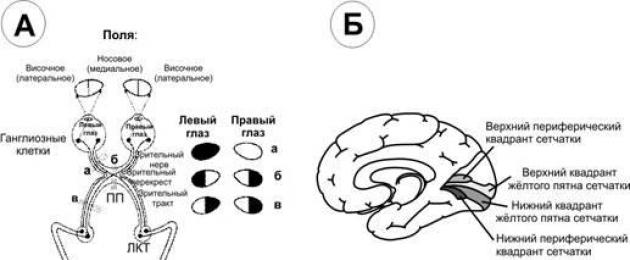
The conductive part of the visual analyzer begins with the optic nerve, which is sent from the orbit to the cranial cavity. In the cavity of the skull, the optic nerves form a partial intersection; Fig. 12.2).
When the coloboma is located in the lower part of the eye, the corresponding visual field deficit is in the upper part. At the same time, when the retina develops in the back of the eye, its front end induces the development of a crystalline lens. This is a vesicle formed by a single layer of cells. in the back of its surface begin to develop in the form of crystalline fibers, organized in a dense and normal way. From a crystalline lens, the tissue undergoes splitting, which leads to the formation of the anterior chamber and the cornea, then the irian tissue between the lens and the cornea, this is the moment when the ciliary body and the iridocorneal.
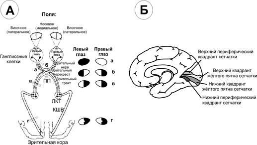
Pic. 12.2. Visuals ways (BUT) and cortical centers (B). BUT. The areas of the optic incision are indicated by lowercase letters, and visual defects occurring after the incision are shown on the right. PP - optic chiasma, LKT - lateral articular body, KSHV - articular-spur fibers. B. The medial surface of the right hemisphere with a projection of the retina in the region of the spore furrow.
The development of the anterior chamber can be disrupted at different stages, and malformations are associated with various structures. The lens may remain attached to the cornea. There may be different crystalline opacities, the iris may be completely missing or incomplete. The pupil may not be in place or have an unusual shape. With iridocorneal angle, sieve structures through which intraocular fluids may not develop, causing glaucoma. The cornea may have a smaller diameter than usual.
The eye itself may be less than normal. The microphthalmic eye can be almost functional, but it often has significant refractive anomalies, corneal or crystalline opacities, and colomatomous changes in the retina and optic nerve. These malformations are an important cause of visual impairment; Therefore, it is important to have a description of this, if possible, in the form of a diagram. The scheme allows you to understand the effect of anatomical malformations on visual function. It is also important to know if there is an optic nerve hypoplasia. that is, if the optic nerve is smaller than normal.
After the intersection, the optic nerves are called the optic tracts. They are directed to the midbrain (to the upper hillocks of the quadrilateral) and the intermediate brain (lateral articulated bodies). The processes of the cells of these parts of the brain as part of the central visual pathway are sent to the occipital region of the cerebral cortex, where the central part of the visual analyzer is located. In connection with an incomplete intersection of fibers, impulses come from the right halves of the retina of both eyes, and to the left hemisphere from the left halves of the retina.
Children with coloboma often have significant refraction errors and therefore need glasses. Children with microphthalmia can also have significant ametropia. Premature infants with coloboma may also have a retinopathy of premature and impaired visual pathways as a result of periventricular leukomalacia. Since the oculomotor pathways are close to these ventricles, these children may have obvious problems with the ocular motor, but sometimes the oculomotor deficiency is also so minimal that they can only be detected by careful neuro-ophthalmologic examination. remember this to plan a sport or physical activity in general.
The structure of the retina. The outermost layer of the retina is formed by the pigment epithelium. The pigment of this layer absorbs light, as a result of which the visual perception becomes clearer, the reflection and scattering of light decreases. To the pigment layer adjacent photoreceptor cells. Because of their characteristic shape, they received the name of rods and cones.
Disability and hearing impairment, communication problems are as varied as visual impairment and visual impairment. It is important to familiarize yourself with the means and level of communication of the child when someone begins to evaluate the visual function. The exam is first held at the usual communication distance; Then you can quickly see the contrast with which the child sees Heidi’s face as a function of distance. Once the connection is established, measurement of visual acuity, visual acuity, contrast sensitivity and color vision becomes easy.
Difficulties in adapting to the dark are rare, but they need to be investigated using photopic and mesoscopic research. Visual field measurements can be taken at the perimeter, campimetry, or methods of confrontation, depending on the level of communication a child has.
Photoreceptor cells on the retina are uneven. The human eye contains 6-7 million cones and 110-125 million rods.
The retina has a 1.5 mm area, which is called blind spot. It does not contain light-sensitive elements at all and is the site of the exit of the optic nerve. 3-4 mm outside of it yellow spotin the center of which there is a small depression central fossa. There are only cones in it, and to the periphery of it the number of cones decreases and the number of rods increases. At the periphery of the retina are only sticks.
Behind the photoreceptor layer is a layer bipolar cells (fig. 12.3), and behind it is a layer ganglion cellsthat are in contact with bipolar. The processes of the ganglion cells form the optic nerve, containing about 1 million fibers. One bipolar neuron is in contact with many photoreceptors, and one ganglion cell with many bipolar cells.
Fig. 12.3. Connection scheme of the receptor elements of the retina with sensory neurons. 1 - photoreceptor cells; 2 –Bipolar cells; 3 - ganglion cell.
From here, it is clear that the pulses from many photoreceptors converge to one ganglion cell, because the number of rods and cones exceeds 130 million. at hit of light on it.
The difference in the functions of rods and cones and the mechanism of photoreception. A number of factors indicate that the rods are a twilight vision apparatus, i.e., they function in twilight, and cones function as a day vision apparatus. Cones perceive rays in bright light conditions. Their activity is related to color perception. The differences in the functions of rods and cones shows the structure of the retina of different animals. Thus, the retina of daytime animals — pigeons, lizards, and others — contains mostly cones, and nocturnal (for example, bats) sticks.
Color is most clearly perceived by the action of rays on the region of the central fossa, but if they fall on the periphery of the retina, then a colorless image appears.
When exposed to rays of light on the outer segment of the sticks, visual pigment rhodopsin decomposed into retinal - a derivative of vitamin A and protein opsin. In the light, after the separation of opsin, retinal is converted directly into vitamin A, which from the outer segments moves into the cells of the pigment layer. Vitamin A is believed to increase the permeability of cell membranes.
In the dark, rhodopsin is restored, for which vitamin A is necessary. When it is deficient, there is a violation of vision in the dark, which is called night blindness. In cones there is a photosensitive substance similar to rhodopsin, it is called iodopsin. It also consists of retinal and opsin protein, but the structure of the latter is not the same as rhodopsin protein.
Due to a number of chemical reactions that take place in photoreceptors, a spreading excitation occurs in the processes of the retinal ganglion cells, which is directed to the visual centers of the brain.
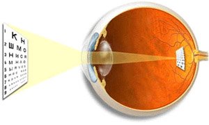
Optical system of the eye. On the way to the photosensitive shell of the eye - the retina - the rays of light pass through several transparent surfaces - the anterior and posterior surfaces of the cornea, lens and vitreous body. Different curvature and refractive indices of these surfaces determine the refraction of light rays inside the eye (Fig. 12.4).
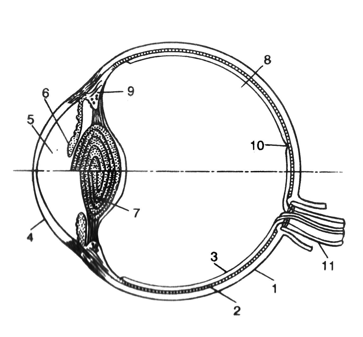
Fig. 12.4. Accommodation mechanism (according to Helmholtz).1 - sclera; 2 - choroid; 3 - the retina; 4 - the cornea; 5 - front camera; 6 - iris; 7 - the lens; 8 - vitreous body; 9 - ciliary muscle, ciliary processes and ciliary belt (cinnamon ligaments); 10 - central fossa; 11 - optic nerve.
The refractive power of any optical system is expressed in diopters (D). One diopter is equal to the refractive power of a lens with a focal length of 100 cm. The refractive power of a human eye is 59 D when viewing distant objects and 70.5 D when viewing close objects. The image obtained on the retina is sharply reduced, turned upside down and from right to left (fig. 12.5).
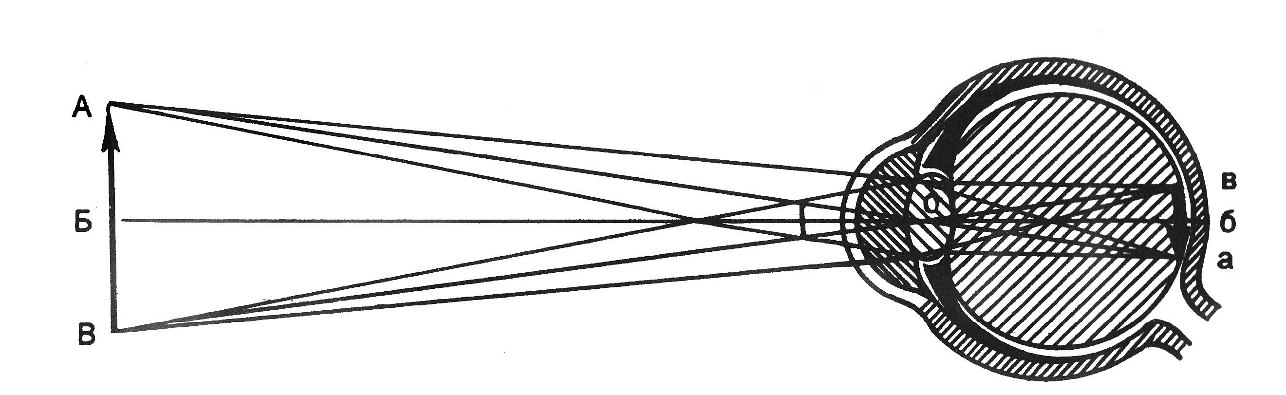
Fig. 12.5. The course of the rays from the object and the construction of the image on the retina of the eye. AB - thing; av - his election; 0 - nodal point; B - b - the main optical axis.
Accommodation. Accommodation called the adaptation of the eye to a clear vision of objects located at different distances from a person. For a clear vision of the object, it is necessary that it be focused on the retina, i.e., that the rays from all points of its surface be projected onto the retinal surface (Fig. 12.6).
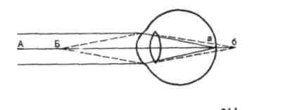
Fig. 12.6. The course of the rays from near and far points.Explanation in the text
When we look at distant objects (A), their image (a) is focused on the retina and they are clearly visible. But the image (b) of close objects (B) at the same time is vague, since the rays from them are collected behind the retina. The main role in accommodation is played by the lens, which changes its curvature and, consequently, its refractive power. When viewing close objects, the lens becomes more convex (Fig. 12.4), thanks to which the rays diverging from any point of the object converge on the retina.
Accommodation is due to the contraction of the ciliary muscles, which alter the convexity of the lens. The lens is enclosed in a thin transparent capsule, which is always stretched, that is, flattened, the fibers of the ciliary belt (Zinn bundle). Contraction of the smooth muscle cells of the ciliary body reduces the craving of the Zinn ligaments, which increases the convexity of the lens due to its elasticity. The ciliary muscles are innervated by the parasympathetic fibers of the oculomotor nerve. The introduction of atropine into the eye causes a disturbance in the transmission of excitation to this muscle, restricting the accommodation of the eye when examining close objects. On the contrary, parasympathomimetic substances - pilocarpine and ezerin - cause contraction of this muscle.
The smallest distance from the object to the eye, at which this object is still clearly visible, determines the position near point of clear visionand the greatest distance is far point of clear vision. When the object is located in the near point, accommodation is maximal, and in the far one - accommodation is absent. The nearest point of clear vision is 10 cm.
Presbyopia.The lens loses its elasticity with age, and as the tension of the Zinn ligaments changes, its curvature changes little. Therefore, the nearest point of clear vision is now not at a distance of 10 cm from the eye, but moves away from it. Close objects with poorly visible. This condition is called presbyopia. Older people are forced to use glasses with biconvex lenses.
Anomalies of refraction of the eye. The refractive properties of a normal eye are called by refraction. The eye, without any disturbance of refraction, connects parallel rays in focus on the retina. If parallel rays converge behind the retina, then develops farsightedness. In this case, the person does not see well the closely spaced objects, while those far away are good. If the rays converge in front of the retina, then it develops myopia, or myopia. With such a violation of refraction, a person does not see well-placed objects, and closely spaced objects are good (Fig. 12.7).
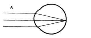
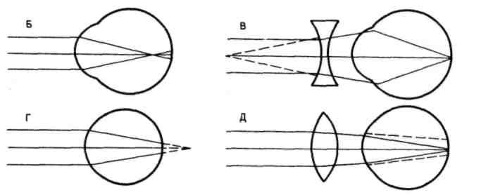
Fig. 12.7. Refraction in normal (A), myopic (B) and long-sighted (D) to the eye and optical correction of myopia (C) and hyperopia (D) scheme
The reason for myopia and hyperopia lies in the unusual size of the eyeball (with myopia, it is elongated, and with hyperopia, it is flattened short) and in an unusual refractive power. When myopia requires glasses with concave glasses that scatter rays; with hyperopia - with biconvex, which collect rays.
Refraction anomalies also apply. astigmatism, i.e., unequal refraction of rays in different directions (for example, along the horizontal and vertical meridians). This deficiency is very weak in every eye. If you look at Figure 12.8, where lines of the same thickness are arranged horizontally and vertically, then some of them seem thinner, others appear thicker.
Fig. 12.8. Astigmatism drawing
Astigmatism is not due to a strictly spherical surface of the cornea. In the case of astigmatism of strong degrees, this surface may approach a cylindrical one, which is corrected by cylindrical lenses that compensate for cornea defects.
Pupil and pupillary reflex. Pupil is the hole in the center of the iris, through which the rays of light pass into the eye. The pupil contributes to the clarity of the image on the retina, passing only the central rays and eliminating the so-called spherical aberration. Spherical aberration is that the rays that fall on the peripheral parts of the lens are refracted more strongly than the central rays. Therefore, if the peripheral rays are not eliminated, the light scattering circles should appear on the retina.
The musculature of the iris is able to change the size of the pupil and thereby regulate the flow of light entering the eye. Changing the diameter of the pupil changes the luminous flux 17 times. The reaction of the pupil to the change in illumination is adaptive, since it somewhat stabilizes the level of illumination of the retina. If you cover your eyes from the light, and then open it, then the pupil that has expanded during an eclipse quickly narrows. This narrowing occurs reflex ("pupillary reflex").
In the iris, there are two types of muscle fibers surrounding the pupil: circular, innervated by the parasympathetic fibers of the oculomotor nerve, the others - radial, innervated by sympathetic nerves. Reduction of the first causes a narrowing, the reduction of the second - the expansion of the pupil. Accordingly, acetylcholine and ezerin cause a narrowing, and adrenaline - the expansion of the pupil. Pupils dilate during pain, during hypoxia, as well as with emotions that increase the excitation of the sympathetic system (fear, rage). Pupil dilation is an important symptom of a number of pathological conditions, such as pain shock, hypoxia. Therefore, dilated pupils with deep anesthesia indicates the onset of hypoxia and is a sign of a life-threatening condition.
In healthy people, the size of the pupils of both eyes is the same. When illuminating one eye, the pupil of the other also narrows; This reaction is called friendly. In some pathological cases, the sizes of the pupils of both eyes are different (anisocoria). This may occur due to the defeat of the sympathetic nerve on the one hand.
Visual adaptation. In the transition from darkness to light, a temporary blindness occurs, and then the sensitivity of the eye gradually decreases. This adaptation of the visual sensory system to bright light conditions is called light adaptation. The reverse phenomenon ( dark adaptation) is observed when moving from a bright room to an almost unlit. At first, a person sees almost nothing due to the reduced excitability of photoreceptors and visual neurons. Gradually, the outlines of objects begin to be detected, and then their details are different, since the sensitivity of photoreceptors and visual neurons in the dark gradually increases.
The increase in light sensitivity during a stay in the dark is uneven: in the first 10 minutes it increases tenfold, and then within an hour - tens of thousands of times. An important role in this process is played by the restoration of visual pigments. Pigments of cones in the dark are restored faster than the rhodopsin of the rods, therefore, in the first minutes of being in the dark, adaptation is due to processes in the cones. This first period of adaptation does not lead to large changes in the sensitivity of the eye, since the absolute sensitivity of the cone apparatus is small.
The next period of adaptation is due to the restoration of rhodopsin rods. This period ends only at the end of the first hour of being in the dark. Restoration of rhodopsin is accompanied by a sharp (100,000 - 200,000 times) increase in the sensitivity of rods to light. Due to the maximum sensitivity in the dark only of rods, a dimly lit object is visible only with peripheral vision.
Theories of color perception. There are several theories of color perception; The three-component theory is most recognized. She claims the existence of three different types of color-sensing photoreceptors in the retina - cones.
The existence of a three-component mechanism of color perception was also mentioned by V.M. Lomonosov. In the future, this theory was formulated in 1801 by T. Jung, and then developed by G. Helmholtz. According to this theory, there are various light-sensitive substances in the cones. Some cones contain a substance that is sensitive to red, others - to green, and others - to violet. Every color has an effect on all three color-sensing elements, but in varying degrees. This theory is directly confirmed in experiments, where the microspectrophotometer measured the absorption of radiation with different wavelengths in single cones of the human retina.
According to another theory proposed by E. Goering, there are substances in cones that are sensitive to white-black, red-green, and yellow-blue radiation. In experiments where a microelectrode was taken away by impulses of ganglion cells of the retina of animals when illuminated with monochromatic light, they found that the discharges of most neurons (dominators) occur under the action of any color. In other ganglion cells (modulators), impulses arise when illuminated with only one color. Identified 7 types of modulators that optimally respond to light with different wavelengths (from 400 to 600 nm).
In the retina and visual centers, many so-called color-optic neurons have been found. The effect of radiation on the eye in some part of the spectrum excites them, and in other parts of the spectrum it slows down. Such neurons are considered to encode color information most efficiently.
Color blindness. Partial color blindness was described at the end of the 18th century. D. Dalton, who himself suffered from it (therefore, the color perception anomaly was called color blindness). Color blindness occurs in 8% of men and is much less common in women: its occurrence is associated with the absence of certain genes in the sexual unpaired X chromosome in men. For the diagnosis of color blindness, important in professional selection, use polychromatic tables. People suffering from this disease cannot be full-fledged drivers of transport, since they cannot distinguish between the color of traffic lights and road signs. There are three types of partial color blindness: protanopia, deuteranopia and tritanopia. Each of them is characterized by a lack of perception of one of the three primary colors.
People suffering from protanopia (“red-blind”) do not perceive red color, blue-blue rays seem to them colorless. People suffering deuteranopia ("Green-blind") do not distinguish green from dark red and blue. With tritanopia - rarely occurring anomalies of color vision, rays of blue and violet are not perceived.
All of these types of partial light blindness are well explained by a three-component theory of color perception. Each type of blindness is the result of the absence of one of the three cone color-sensing substances. There is also full color blindness - achromasiain which, as a result of the defeat of the retina conic apparatus, a person sees all objects only in different shades of gray.
The role of eye movement for vision. When viewing any items, the eyes move. Eye movements are performed by 6 muscles attached to the eyeball. The movements of two eyes are performed simultaneously and amicably. Considering close objects, it is necessary to reduce, and looking at distant objects - to separate the visual axes of two eyes. The important role of eye movements for vision is also determined by the fact that for the brain to continuously receive visual information, an image movement on the retina is necessary. Impulses in the optic nerve occur at the moment of switching on and off the light image. When the light acts on the same photoreceptors, the pulsation in the optic nerve fibers quickly stops and the visual sensation with fixed eyes and objects disappears after 1-2 seconds. To prevent this from happening, the eye, when looking at any object, produces continuous jumps that are not felt by the person. Due to each jump, the image on the retina shifts from one photoreceptor to a new one, again causing impulses of ganglion cells. The duration of each jump is one-hundredth of a second, and its amplitude does not exceed 20º. The more complex the object in question, the more complex the trajectory of eye movement. They, as it were, trace the contours of the image, lingering on its most informative sites (for example, in the face - these are eyes). In addition, the eye continuously finely trembles and drifts (moves slowly from the point of gaze fixation) - the saccades. These movements also play a role in the maladaptation of visual neurons.
Types of eye movements. There are 4 types of eye movements.
Saccades - unsightly quick jumps (in hundredths of a second) eyes, tracing the contours of the image. Saccadic movements contribute to the retention of the image on the retina, which is achieved by periodic displacement of the image along the retina, leading to the activation of new photoreceptors and new ganglion cells.
Smooth follow eye movements behind a moving object.
Converging movement - reducing the visual axes towards each other when viewing an object close to the observer. Each type of movement is controlled by the nervous apparatus separately, but ultimately all mergers end in motor neurons innervating the outer muscles of the eye.
Vestibular eye movements - a regulating mechanism that appears when the receptors of the semicircular canals are excited and that maintains the fixation of the gaze during head movements.
Binocular vision. When looking at any object, a person with normal vision does not have the sensation of two objects, although there are two images on two retinas. The images of all objects fall on the so-called corresponding, or corresponding, sections of two retinas, and in human perception these two images merge into one. Press down slightly on one eye from the side: it will immediately begin to split in the eyes, because the retina matching is broken. If you look at a close object, converging eyes, then the image of any more distant point falls on the nonidentical (disparate) points of two retinas (Fig. 12.9). The disparate plays a large role in estimating the distance, and therefore in the vision of the depth of the relief. A person is able to notice a change in depth, creating a shift of the image on the retinas by several angular seconds. Binocular fusion or integration of signals from two retinas into a single visual image occurs in the primary visual cortex. Vision with two eyes greatly facilitates the perception of space and the depth of the object, contributes to the definition of its shape and volume.
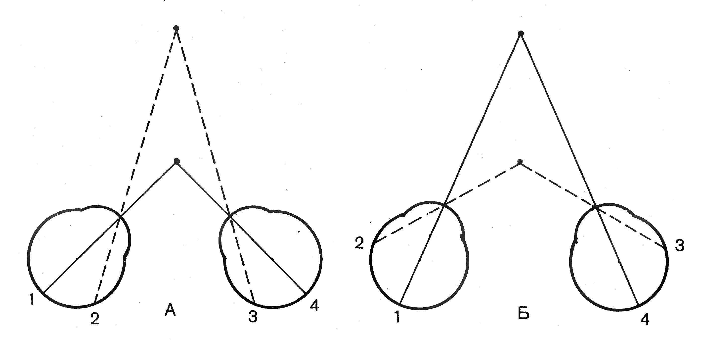
Fig. 12.9. The course of the rays with binocular vision. BUT - fixing the eyes of the nearest subject; B - fixing gaze distant subject; 1 , 4 - identical retinal points; 2 , 3 - non-identical (disparate) points.
More than 80% of the information we receive with our eyes. The structure of the eye is extremely complex and depends on the functions it performs.
____________________________
The structure of the human eye
The constituent parts of the human eye as a paired organ of vision are:
- eyeball,
- optic nerve
- tear glands
- eyelid,
- muscles of the eyeball.
The eyeball of man and other higher animals- This is a sphere of irregular shape, with a diameter of 2.5 cm. Two eyeballs are located inside the orbits (eye cavities) of the skull. It is noteworthy that the eyeballs of different people differ approximately in fractions of a millimeter. From the moment of birth until the death of the individual, the eye sockets are doubled.
An important part of the structure of the human eye is the optic nerve, With the help of which information about the object is transmitted to the occipital cortex, where it is analyzed.
In the structure of the eye, the scheme of which is shown, an important role is played subsidiary bodies. Thanks tear glandthat is located in the upper part of the orbit of the eye, the surface always remains wet. A tear lubricates the conjunctiva well and has a bactericidal effect due to the lysozyme enzyme present in it. The performance of optical functions is possible due to the fact that the eye is moistened. Human lacrimal glands secrete about 0.5-1 ml of secretion per day, which means 25 liters in a lifetime.
The upper and inner eyelid covers the eye, protecting it from negative environmental factors.The same function is performed by the eyelashes, which grow on the edge of the eyelids. The structure of the human eye is such that it ensures the coordinated action of the six muscles of the eyeball.
Important elements that includes the structure of the human eyeball
The eyeball consists of three shells that surround the transparent contents of the eye:
- vitreous body
- lens,
- intraocular fluid front and rear cameras.
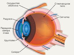
The outer membrane of the sclera (protein)- consists of rigid and fibrous tissue that protects the eye from mechanical damage. It provides the shape and volume of the eye. The white color of the sclera contrasts with the iris. The anterior transparent area is the cornea, behind which the anterior chamber is located.
In the structure of the eye, the scheme of which is on the site, it is clear that a thin iris is located behind the cornea. Different people have it differently. Brown eye color is considered the most common on the planet, whereas only 2% of people on Earth can boast of green iris. The color of a person’s eyes depends on the amount of melanin in the body (brown-eyed people have a lot of it). On the retina are the sensitive cells (photoreceptors) and blood vessels that feed them.
The presentation “The structure of the eye” shows that the most sensitive place of the retina is the “yellow spot” zone, where are millions of tightly packed photoreceptors (cones). The high density of cones in the "yellow spot" creates a very detailed picture, like a high-resolution digital camera with a large number of megapixels. Each photoreceptor is associated with nerve fibers that collectively form the optic nerve.
There are two main types of photoreceptors:
- cones (responsible for detailed central vision),
- sticks (responsible for night vision and peripheral vision).
Photoreceptors in the retina convert the image into electrical signalsthat enter the brain through the optic nerve. In the structure of the eye, the pictures clearly illustrate the division of the eyeball into two chambers, each of which is filled with liquid. The anterior chamber consists of an intraocular fluid that feeds internal structures. The back chamber consists of a gelatinous liquid (vitreous body), which helps to create pressure inside the eye to maintain its shape.
The relationship of the structure and complex functions of the human eye
To understand how this complex organ functions, you need to consider the structure of the human eye, pictures of which describe in detail all the components.
It is believed that the eye is a rather imperfect optical system. The best way to understand the structure and function of the eye is to compare it with a camera. The camera creates an image by focusing on the subject and allowing a specific amount of light to pass through the aperture of the aperture. The structure of the eye is such that it performs its functions in a similar way.
When light enters the eye, it passes through the cornea (lens).where 2/3 of the light focusing is achieved. The smallest curvature changes allow the cornea to substantially focus the light beam. Then the light hits the pupil, where its constriction or expansion, like the diaphragm, regulates the amount of light. The lens is the second strong lens of the eye, which provides 1/3 of the focusing of the light beam.
The shape of the lens can be changed by tension or relaxation of the muscles of the eye. The focused light beam reaches the retina, where it is converted into a nerve impulse. When the image reaches the brain centers, we are able to enjoy the beauty of the world, we see colors, objects and are able to react to danger in time. Thus, the structure and function of the eye are in a clear relationship, representing an amazing evolutionary masterpiece of the human body.
The structure of the eye - the subject of studying scientists from different fields of knowledge for a dozen centuries. Physiologists, neuroscientists, biophysicists and ophthalmologists argue about the origin and functioning of the organs of vision. They agree only that the shape of the human eye is optimal in order to exchange views and attract other individuals.
The presentation of the structure of the eye shows how complex and amazing our eyes are.Doctors are still unable to find a way to transplant the eyeballs, since the optic nerve is extremely complex and sensitive and cannot be successfully recovered. The proverb says that the valuable thing must be kept as the pupil of the eye. This emphasizes the importance and indispensability of vision for a person.
The structure and work of the human eye, video
- In contact with 0
- Google+ 0
- OK 0
- Facebook 0