The retina is the initial part of the visual analyzer, which provides for the perception of light waves, their transformation into nerve impulses and transmission into the optic nerve. Photoreception is one of the most important and complex processes that allow a person to see the world around.
Today, retinal pathology is an actual problem of ophthalmology. Diabetic retinopathy, acute obstruction of the central artery, various detachments and retinas are common causes of irreversible blindness in developed countries.
With anomalies of the structure of the retina are associated, night blindness (poor lighting of the room prevents a person from seeing normally) and some other visual disorders. Knowledge of the anatomy and physiology of the retina is necessary for understanding the mechanism of development of pathological processes in it, the principles of their treatment and prevention.
What is the retina
The retina is the inner lining of the eye lining the inside of the eyeball. Knutri from it is the vitreous body, outwards - the choroid. Retina is very thin - normally its thickness is only 281 microns. It should be noted that in the area of the macula it is slightly thinner than at the periphery. Its area is about 1206 mm 2.
The reticular membrane lines approximately ¾ of the inner surface of the eyeball. It stretches from the optic nerve head to the dentate line, where it enters the pigment epithelium and lines the inside of the ciliary body and the iris. In the dentate line and the optic disc, the retina is attached very firmly, in all other places it is loosely connected to the pigment epithelium, which separates it from the choroid. It is the absence of a tight connection that makes the development of retinal detachments so easy.
The layers of the retina are unequal in structure and function, and together they form a complex structure. It is thanks to the close contact and interaction of various parts of the visual analyzer that people are able to distinguish colors, see surrounding objects and determine their sizes, estimate distances, and perceive the surrounding world adequately.
Getting into the eye, the incoming rays pass through all its refracting media - the cornea, chamber moisture, lens, vitreous body. Due to this, in people with normal refraction, the image of surrounding objects is focused on the retina - reduced and inverted. Further, the light pulses are transformed and enter the brain, where the picture that a person sees is formed.
Functions
The main function of the retina is photoreception - a chain of biochemical reactions, during which light stimuli are converted into nerve impulses. This is due to the breakdown of rhodopsin and iodopsin - visual pigments that form when there is enough vitamin A in the body.
The reticular membrane of the eye provides:
- Central vision . It allows a person to read, perform work close up, and clearly see objects located at different distances. The retinal cones, which are located in the area of the macula, are responsible for it.
- Peripheral vision . Necessary for orientation in space. It is provided by sticks, which are localized paracentrally and on the periphery of the retina.
- Color vision . It makes it possible to distinguish colors and their shades. Three different types of cones are responsible for it, each of which perceives light waves of a certain length. This enables a person to distinguish between green, red and blue colors. Violation of color perception is called color blindness. Some people have such a phenomenon as the fourth, extra cone. It is typical for 2% of women who can distinguish up to 100 million colors.
- Night vision . Provides the ability to see in low light conditions. It is carried out thanks to chopsticks, since cones in the dark do not function.
Retinal structure
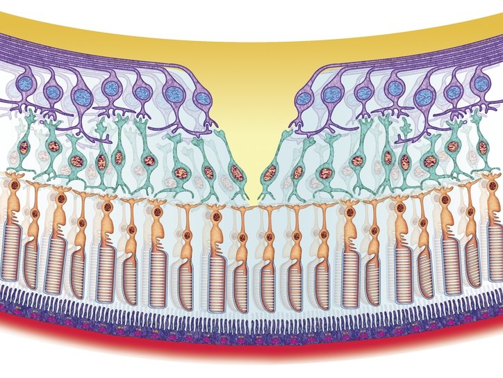
The structure of the retina is very complex. All its elements are closely related, and damage to any of them can lead to serious consequences. Retina has a three-neuronal receptor-conducting network necessary for visual perception. This network consists of photoreceptors, bipolar neurons and ganglion cells.
Retinal layers:
- Pigment epithelium and Bruch's membrane . Perform barrier, transport, trophic functions, prevent the penetration of light radiation, phagocytic (absorb) segments of rods and cones. In some diseases, hard or soft druses are formed in this layer - small spots of yellow-white color. .
- Photosensory layer . It contains retinal receptors, which are outgrowths of photoreceptors - highly specialized neuroepithelial cells. Each photoreceptor contains a visual pigment that absorbs light waves of a certain length. Rods contain rhodopsin, cones contain iodopsin.
- Outer boundary membrane . Formed by terminal plates and flat adhesive contacts of photoreceptors. Also here are located the external processes of the Mullerian cells. The latter perform the light guide function - they collect light on the anterior surface of the retina and conduct it to the photoreceptors.
- Outer nuclear layer . It contains the photoreceptors themselves, namely, their bodies and nuclei. Their external processes (dendrites) are directed in the direction of the pigment epithelium, and internal - to the outer network layer, where they come into contact with bipolar cells.
- Outer mesh layer . Formed by intercellular contacts (synapses) between photoreceptors, bipolar cells and associative neurons of the retina.
- Inner nuclear layer . Here lie the bodies of Mullerian, bipolar, amacrine and horizontal cells. The former are cells of the neuroglia and are necessary for the maintenance of nervous tissue. All others process the signals coming from photoreceptors.
- Inner mesh layer . Contains internal processes (axons) of various nerve cells of the retina.
- Ganglion cells receive impulses from photoreceptors through bipolar neurons, and then conduct them to the optic nerve. These nerve cells are not covered with myelin, due to which they are completely transparent and easily transmit light.
- Nerve fibers . They are axons of ganglion cells that transmit information directly to the optic nerve.
- Inner boundary membrane . Separates the retina from the vitreous body.
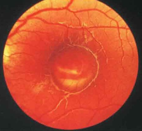
A little medial (closer to the middle) and up from the center of the retina in the fundus of the eye is the optic nerve head. It has a diameter of 1.5-2 mm, pink color, and in its center there is noticeable physiological excavation - a notch of small size. In the area of the ONH is a blind spot, devoid of photoreceptors and insensitive to light. In determining the visual fields, it is defined in the form of a physiological scotoma - the loss of part of the visual field.
In the central part of the optic nerve head there is a small depression through which the central artery and the vein of the retina pass. Retinal vessels lie in the layer of nerve fibers.
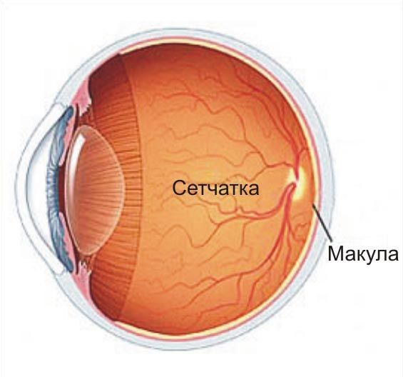
Approximately 3 mm lateral (closer to the outside) of the optic disc, there is a yellow spot. In its center is located the central fossa - the location of the largest number of cones. It is she who is responsible for high visual acuity. Pathology of the retina in this area has the most adverse effects.
Methods of diagnosis of diseases
The standard diagnostic program includes measuring intraocular pressure, checking visual acuity, determining refraction, measuring visual fields (perimetry, campimetry), biomicroscopy, direct and indirect ophthalmoscopy.
Diagnostics may include the following methods:
- study of contrast sensitivity, color perception, color thresholds;
- electrophysiological diagnostic methods (optical coherence tomography);
- fluorescein angiography of the retina - allows you to assess the condition of the vessels;
- photographing the fundus - necessary for further observation and comparison.
Symptoms of retinal diseases
The most characteristic sign of lesion of the retina is the reduction of sharpness or narrowing of the visual fields. It is also possible the appearance of absolute or relative livestock of different localization. Various forms of color blindness and night blindness may indicate a photoreceptor defect.
A marked deterioration in central vision indicates a lesion of the macular region, and the peripheral one - the periphery of the fundus. The appearance of scotoma suggests local damage to a specific zone of the retina. The increase in the size of the blind spot, along with a strong decrease in visual acuity, can speak about the pathology of the optic nerve.
Occlusion of the central retinal artery is manifested by sudden and abrupt (within a few seconds) blindness of one eye. With tears and detachments of the retina, the appearance of light flashes, lightning, glare before eyes is possible. The patient may complain about the occurrence of fog, black or colored spots in the field of view.
Retinal diseases
According to etiology and pathogenesis, all diseases of the retina are divided into several large groups:
- vascular disorders;
- inflammatory;
- dystrophic lesions;
- injuries;
- benign and malignant neoplasms.
The treatment of each disease of the retina has its own characteristics.
To combat the pathological changes of the retina can be used:
- anticoagulants - Heparin, Fraxiparin;
- retinoprotectors - Emoxipin;
- angioprotectors - Ditsinon, Troxevasin;
- vasodilators - Sermion, Cavinton;
- vitamins of group B, nicotinic acid.
The drugs are administered parabulbarno (eye injections), less commonly used eye drops. During ruptures, detachments, and severe retinopathies, laser coagulation, circlages, episcleral filling, cryopexy can be performed.
Inflammatory diseases are retinitis of various etiologies. Inflammation of the retina develops due to the ingress of microbes into it. If everything is simple, then you should tell more about other groups of diseases.
Vascular pathology
One of the most frequent vascular diseases of the retina is the defeat of vessels of different caliber. The cause of its development can be hypertension, diabetes, atherosclerosis, trauma, vasculitis, osteochondrosis of the cervical spine.
Initially, patients may have dystonia or angiospasm of the retina, later hypertrophy, fibrosis or vascular thinning develops. This leads to ischemia of the retina, which causes angioretinopathy in the patient. In individuals with hypertension, an arterio-venous cross appears, symptoms of copper and silver wire. Diabetic retinopathy is characterized by intense neovascularization, a pathological proliferation of blood vessels.
Retinal angiodystonia is manifested by a decrease in visual acuity, flashing flies before the eyes and visual fatigue. Arterospasm can occur with elevated or lowered arterial pressure, some neurological disorders. In parallel with the defeat of the arterial vessels, the patient may develop phlebopathy.
A common vascular pathology is occlusion of the central retinal artery (OCAC). The disease is characterized by blockage of the vessel or one of its branches, leading to severe ischemia. Embolism of the central artery most often occurs in individuals with atherosclerosis, hypertension, arrhythmia, neurocirculatory dystonia and certain other diseases. Treatment of pathology should begin as soon as possible. If medical care is not provided in time, occlusion of the central retinal artery can lead to complete loss of vision.
Dystrophies, injuries, malformations
One of the most common malformations is the coloboma - the absence of a portion of the retina. Often there are macular (mainly in the elderly), central, peripheral. The latter are divided into different types: lattice, small cystic, frostlike, “snail track”, “cobblestone pavement”. With these diseases in the fundus you can see defects resembling holes of different sizes. Pigmented degeneration of the retina is also found (its cause is redistribution of the pigment).
After blunt traumas and contusions, Berlin turbidity often appears on the retina. Treatment of pathology is the use of antihypoxants, vitamin complexes. Often, sessions of hyperbaric oxygenation are prescribed. Unfortunately, the treatment does not always have the expected effect.
Neoplasm
The retinal tumor is a relatively frequent ophthalmic pathology - it is 1/3 of all neoplasms of the eyeball. Usually, patients are diagnosed with retinoblastoma. Nevus, angioma, astrocytic hamartoma and other benign neoplasms are less common. Angiomatosis is most often combined with various malformations. Tactics of treating neoplasms is determined individually.
The retina is the peripheral part of the visual analyzer. It carries out photoreception - the perception of light waves of various lengths, their transformation into a nerve impulse and conducting it to the optic nerve. With retinal lesions, people experience a wide variety of visual disturbances. The most dangerous consequence of retinal damage is blindness.
Pathological conditions of the retina and optic nerve are often predetermined by cardiovascular, neurological and other diseases, as well as endocrine disorders, which necessitate a general coordinated treatment of such patients by an ophthalmologist and a doctor of the relevant specialty. In addition, changes in the fundus attached great diagnostic and prognostic value.
It should be especially noted that retinal diseases, primarily vascular and dystrophic lesions, are today one of the main causes of blindness and visual disability, which indicates the need for early diagnosis and timely complex treatment of both an ophthalmologist and general practitioners.
Retinal Anatomy
Retina(retina) peripheral part of the visual analyzer. It develops from the front of the brain bladder, because it can be considered part of the brain, carried to the periphery. It distinguishes 10 layers: 1) a layer of pigment epithelium; 2) a layer of rods and cones; 3) outer boundary membrane; 4) outer nuclear layer; 5) the outer reticular layer; 6) inner nuclear layer; 7) the inner reticular layer; 8) a layer of multipolar (ganglion) cells; 9) a layer of nerve fibers; 10) inner boundary membrane. There are 3 specific visual neurons in the retina:
1. Sticks and cones ( cellula optіca bacіllіformіs et conіformіs).
2. Bipolar cells ( neurocytus blue polaris).
3. Ganglion cells ( neurocytus ganglіonarіs).
The rods have a very high light sensitivity, provide twilight and peripheral vision, there are a lot of them (about 130 million), they are located along the entire periphery of the retina to the border of its optical part ( ora serrata).
Cones are located mainly in the area of the central fossa of the yellow spot, there are about 7 million of them. They provide a uniform vision and color perception.
The first neuron lies on the layer of the pigment epithelium, firmly associated with the choroid, which ensures the continuous restoration of the molecules of the visual pigments (rhodopsin and iodopsin) necessary for the photochemical process of the act of vision. Thus, the function of the retina is closely related to the condition of the choroid itself.
The second neuron is associative.
The third neuron has long processes that form the optic nerve.
The internuclear retinal layers consist of fibrous structures and form the core of the retina. The processes of the ganglion cells form the optic nerve, which exits the orbit through the optic hole. In the middle cranial fossa, in the region of the Turkish saddle, a partial intersection of the optic nerve fibers of both eyes occurs (only the medial fibers intersect). After the intersection of the so-called optic tract, which contains fibers from the retina of both eyes. The subcortical center of the visual analyzer is the external articulated bodies, and the cortical орная is the spur-like groove in the occipital lobe of the brain ( fіssura calcarina).
The blood supply of the retina is carried out from the central artery of the retina, the trophicity of its outer sections is provided by the choriocapillary layer of the choroid. The retina has no sensitive innervation, so the defeat of it does not cause pain.
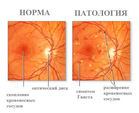
The normal fundus of the eye has the following form: the optic nerve disc is pink, its boundaries are clear, the arteries and veins of the retina are of a uniform caliber, the ratio of the caliber of the artery to the caliber of the vein is 2: 3, there are no focal changes.
Diagnostics retinal diseases based on ophthalmoscopy, fluorescence angiography, functional and electrophysiological studies (visual acuity, visual field, color perception, dark and light adaptation, electroretinography, electrical sensitivity of the optic nerve for phosphene, optical coherent tomography).
Complaints of patients are non-specific and consist in dysfunction of central vision (photopsia, metamorphosis, reduced visual acuity, central scotomas, disturbed color sensation) or peripheral vision (restriction and loss in sight, reduction of dark adaptation).
Ophthalmoscopic changes may be as follows:
1. Change of gauge, walls and stroke of vessels.
2. Hemorrhages of various shapes, sizes and abundances.
3. Diffuse or local opacities of the retina (foci).
4. Pigment deposits (foci, mottling).
Pathology of the retina is extremely diverse. Among the diseases of the retina are the following main forms:
1. Diseases associated with common diseases of the body.
2. Inflammatory diseases.
3. Dystrophic changes.
4. Retinal detachment.
5. New growths.
6. Anomalies of development.
Let's stop on those diseases of a retina which most often meet, doctors of all specialties have to be familiar with them.
Cardiovascular diseases lead to various changes in the fundus. So, in hypertension, these changes reflect the pathogenesis of vascular disorders that occur in the body, and are of great diagnostic and prognostic value. According to the classification A.Ya. Vilenkina, MM Krasnov, distinguish: hypertensive angiopathy, hypertensive angiosclerosis, hypertensive retinopathy, hypertensive neuroretinopathy.
With hypertensive angiopathythere are expansion, tortuous veins, narrowing of the arteries, their uneven caliber. Observed with stage I of hypertension.
With hypertensive angiosclerosis in addition to the above described phenomena of angiopathy, an uneven light reflex, symptoms of copper and silver wire, a symptom of arteriovenous chiasm (Salius-Gunn I, II and III degrees) appear along the thickened walls of the arteries.
Symptom Salus-Gunn I: conical narrowing of the vein on both sides of the artery at the place of their intersection, the vein takes the form of an hourglass. Symptom Salus-Gunn II: in the place of an arteriovenous junction, the vein is arcuately bent and pushed back into the retina. Symptom Salus-Gunn III: a vein in the place of intersection can not be distinguished, as it is covered by the edematous retina. This phenomenon is characteristic of stage II and III stages of hypertension.
With hypertensive retinopathy there are foci and hemorrhages in the retina, reduced vision. Observed with stage III hypertension.
Hypertensive neuroretinopathy adverse prognostic sign. The process involves the optic nerve. There is edema of the optic nerve head, hemorrhages and retinal edema appear around it. Visual acuity decreases, the field of view narrows. Observed with stage III hypertension.
However, there may not be a complete parallel between the clinical course of hypertension and the fundus picture.
Treatment. They treat the underlying disease. In retinopathy, in addition, resorption therapy is used (fibrinolysin, parabulbarny hemase), angioprotectors, antioxidants (emoxipin, ditsinon, doksium), with neuroretinopathy моч also diuretic and osmotic agents.
With renal hypertensionnarrowing of the arteries, dilatation of the veins of the retina without marked sclerotic changes, with a large number of exudative foci and plasmorrhagia. A typical is the figure of a star in the macular region. This is a bad prognostic sign, in the words of old authors "death knell" for the patient. It was previously believed that life expectancy with the appearance of such changes in the fundus was 1-3 years, but now, thanks to effective treatment, in many cases it is possible to achieve a significant improvement in the general condition of the patient with a complete or partial reverse development of hypertensive changes in the fundus.
Diabetes mellitus is a common cause of severe retinal lesions, which are called diabetic retinopathy. They consist in the appearance of microaneurysms, hemorrhages, exudative foci; in the terminal phase the development of proliferating processes, the emergence of newly formed vessels, proliferation of connective tissue, the development of secondary retinal detachment.
Treatment is the use of angioprotectors, absorbable agents, anabolic hormones. In recent years, photo- and laser coagulation and cryotherapy have been used. The prognosis is unfavorable.
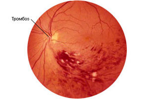
The general vascular pathology of the organism leads to the development of such diseases of the retina as acute obstruction of the central retinal artery, and thrombosis of its central vein.
Obstruction of the central retinal artery caused by spasm (50%), thrombosis (45%) or embolism (5%) of the artery. It occurs, in addition to patients with hypertension, in young persons suffering from endocarditis, in particular, rheumatic, chronic infectious diseases.
Patients complain of sudden loss of vision, up to light perception. In the fundus determine the sharp narrowing of the arteries, edema of the retina, the symptom of “cherry seed”. As a result of the disease, atrophy of the optic nerve develops.
Treatment: vasodilators (0.1% solution of atropine retrobulbar, intravenous nicotinic acid, aminophylline, trental; sublingual nitroglycerin), thrombolytic agents, anticoagulants.
Forecast unfavorable. Treatment is effective when treated in the first 2-4 hours after the disease.
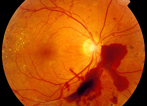
Thrombosis of the central retinal vein occurs mainly in the elderly, suffering from hypertension, atherosclerosis. Patients complain of a sudden sharp decrease in vision, but complete blindness does not happen. Multiple hemorrhages, plasmorrhages, dilation and tortuosity of the veins, discontinuity of their course, edema of the retina, and thinning of the boundaries of the optic nerve head (the so-called "crushed tomato" symptom) are visible on the fundus of the eye.
Prognosis for vision bad, but more favorable than in case of obstruction of the central retinal artery. After resorption of hemorrhages, atrophic foci form in the retina, in some patients secondary glaucoma develops.
Treatment: anticoagulants of direct and indirect action, thrombolytic and absorbable drugs.
Inflammatory diseases of the retina
These include metastatic retinitis, chorioretinitis. They arise as a result of contact with the blood flow of microorganisms from any suppurative focus.
Complaints of the patient depend on the localization process. Lesions of the central parts of the retina are accompanied by metamorphosis, decreased visual acuity, the appearance of cattle, with peripheral localization of foci complaints may be absent.
Diagnosis set with ophthalmoscopy. On the fundus visible yellowish-white lesions with fuzzy boundaries that rise above the retina, over time atrophic chorioretinal lesions develop in their place.
Treatment: anti-inflammatory and resorption therapies, a comprehensive examination of the patient to establish the etiology of the disease.
Dystrophic retinal changes
There are the following types of degenerative changes in the retina:
1. Hereditary generalized dystrophies (pigmentary dystrophy of the retina, congenital Leuro amaurosis).
2. Hereditary peripheral dystrophy of the retina.
3. Hereditary central retinal dystrophy.
4. Age-related retinal dystrophy.
Pigmentary dystrophy of the retina (PDS). The disease is familial with hereditary recessive inheritance.
Complaints of patients: decrease and loss of vision at dusk (hemelopia), then narrowing of the visual field develops, in the terminal stage остр visual acuity decreases, up to complete blindness.
When the PDS in the fundus appear, starting from the periphery, the pigment lesions in the form of bone bodies, which subsequently capture and central regions. Retinal vessels narrow sharply. The optic nerve head becomes pale, with a waxy tinge; its complete atrophy develops at the terminal stage. The prognosis is unfavorable.
Treatment: vasodilators, metabolic drugs, vitamins, tissue therapy, hormones, anabolic steroids, revascularization operations, retrosclerosis, physiotherapeutic treatment (ultrasound, phonophoresis, electrophoresis, electrostimulation by “phosphene”, magnetic therapy).
Hereditary macular dystrophy.There are a large number of clinical forms that differ in the pattern of the fundus and the nature of the clinical course.
Diseases are family-hereditary in nature, are transmitted by the recessive or dominant type and are distinguished by a steadily progressive course. Dystrophy of the yellow spot appears in preschool or school, sometimes in adolescence. It should be remembered that macula degeneration in children is also observed in the first year of life in case of Tay-Sachs, Niemann-Pick diseases.
Tay-Sachs disease (familial amaurotic idiocy) is characterized by blindness with typical changes in the yellow spot (a grayish-white focus with a “cherry bone” in the center), strabismus and nystagmus, mental retardation up to complete dementia, progressive muscle weakness. Fatal outcome is up to two years.
In Niemann-Pick disease (reticuloendothelial sphingomyelinosis), a grayish-white focus with a "cherry bone" in the macula, yellowish atrophic optic nerve head, exophthalmos, nystagmus, enlargement of the liver, spleen, and mental and physical development are characteristic. Fatal outcome is up to two years.
Age-related retinal dystrophyare peripheral and central. Peripheral dystrophies can lead to tearing and retinal detachment. Prophylactic conduct cryopexy, laser coagulation.
Macular dystrophies are extremely common, according to various authors, their incidence among persons over 50 is 15 29%. Patients complain of a gradual decrease in vision, as a result of which vision decreases to a hundredth, a central absolute scotoma appears.
Two forms of sclerotic macular dystrophy are clinically distinguished: “dry” and exudative-hemorrhagic. When the “dry” form on the fundus of the eye has atherosclerotic changes in the retinal vessels, deposits in the retina of lipids, cholesterol, hyaline (drusen), depigmentation, atrophic lesions.
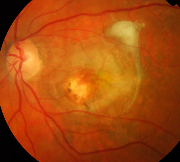
When the exudative hemorrhagic course of the disease in the fundus appears yellowish-white discoid lesion, surrounded by hemorrhages. Subsequently, the lesion will replenish into the vitreous, so it must be differentiated from the choroid neoplasm (melanoblastoma) - this is the so-called pseudotumorosis nidus.
Treatment: in case of “dry” macular dystrophy physiotherapeutic methods of treatment, vitamin therapy, metabolites, vasodilators, antioxidants, revascularization operations, temporal artery ligation, retrosclerosis. When edematous form angioprotectors, antioxidants, resorption therapy, laser coagulation, cryopexy.
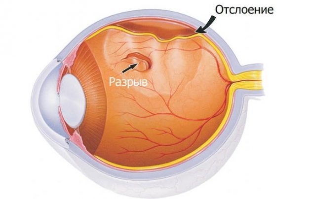
Retinal dystrophy is a factor that can lead to development retinal detachment, especially when stretching the eye (with high myopia). Retinal detachment may also occur under the influence of cicatricial changes in the vitreous body. Most often, the direct cause of it is injury or physical stress. The development of retinal detachment is due to the fact that the retina is anatomically closely connected with the underlying tissue in only two places: near the dentate line in the flat part of the ciliary body and near the optic nerve head.
Patients complain of the appearance of flashes of light or "lightning" (photopsies) on the periphery of the visual field in the area opposite to the retinal break. Then a sensation of a “veil” appears, which comes from the same side, from the periphery of the visual field to its center, the narrowing of the visual field occurs, most often from above.
When ophthalmoscopy area of detachment has the appearance of a bubble or sail grayish color, against which the retinal vessels look dark, and the breaks bright red.
Before hospitalization, such a patient should be provided with bed rest, preferably with a binocular bandage. Emergency hospitalization is indicated.
Treatment surgical. An operation is performed on depressions of the sclera with diathermocoagulation or cryopexy for the development of scar tissue that holds the detached retina. In recent years, laser coagulation, as well as intravitreal surgical interventions have been widely used in the treatment of retinal detachment. In these operations, vitrectomy is performed (removal of the altered vitreous body, vitreoretinal moorings and proliferative epiretinal membranes). In order to flatten the retina to the choroid, gases that expand (organofluorinated compounds) or silicone oil are injected. If necessary, dissection of the shortened detached retina is performed and straightened with fixation of the edges using cryo- or endolaser coagulation. In some cases, microscopic retinal nails and magnets are used.
Retinoblastoma (glioma)Malignant neoplasm of the retina, which occurs in the first months or years of a child’s life. In its course there are 4 stages.
Stage I initial. The limited tumor site in the retina is determined.
Stage II germination in the eye cavity, in the angle of the anterior chamber. The characteristic symptom is ам "amarotic cat's eye", the pupil is dilated, yellowish, intraocular pressure is increased.
Stage III germination of the tumor into orbit. Exophthalmos may appear. The tumor is growing rapidly anteriorly, has the appearance of cauliflower.
Stage IV metastasis to distant organs, germination in the cranial cavity.
The differential diagnosis is carried out with retrolental fibroplasia, in which intraocular pressure is normal, as well as with the outcome of purulent septic endophthalmitis, which is usually accompanied by hypotonia of the eye. The most informative are the following diagnostic methods: ultrasound, computed tomography, radioisotope research. Diaphanoscopy is less informative.
Treatment: in the І and ІІ stages enucleation; in ІІІ and ІV exentation of the orbit with subsequent x-ray and chemotherapy.
Forecastadverse.
Retinopathy of prematurity
According to modern concepts, retinopathy (PH) is a vascular proliferative lesion of the retina, which occurs predominantly in premature babies and, in some cases, leads to irreversible blindness.
PH occurs, on average, in 20% of premature babies, 5–7% of them pathological changes lead to complete loss of vision.
It has been proven that PH develops in somatically, neurologically and perinatally complicated children. However, low birth weight (1500g and less), gestational age at birth 32 weeks or less, as well as oxygen therapy, which takes more than 30 days, are the most significant and constant risk factors.
With PH, the active phase and the regression phase are distinguished. The active phase is divided into five stages:
І stage - on the border of the vascularized and avascular areas of the retina, a demarcation line of white color is formed, which is an accumulation of vessel-forming mesenchymal tissue.
Stage II - a protrusion is formed in the area of the demarcation line, which may acquire a pink color as a result of intraretinal neovascularization. The protrusion begins to flash above the level of the retina.
Stage III- in the place of the protrusion, fibrous vascular extraretinal proliferation is formed.
IV stage incomplete retinal detachment.
Stage V - total retinal detachment.
At present, it is generally accepted that there are no sufficiently effective methods of conservative treatment of PH (World Forum of Pediatric Ophthalmologists, London, 2000).
Surgical treatments for PH include cryotherapy, laser photocoagulation and lensvitrectomy combined with scleral filling.
Requirements for the work of a neonatologist and an ophthalmologist are the need to inform parents of premature newborns, especially those with low and extremely low birth weight, in a timely manner about the potential for the occurrence and severity of PH, the potential seriousness of this disease and the importance of timely ophthalmologic studies.
The retina (retina) is one of the most complex structures of the visual organ. She is responsible for the projection of images and their transmission in the form of electrical impulses through the optic nerve to the brain. Therefore, any diseases of the retina lead primarily to the deterioration of vision. In order to preserve visual ability and to avoid radical measures for its recovery, it is important to promptly identify diseases and treat them at an early stage. But today there are about ten pathologies of the retina: how to distinguish them from each other?
The retina is a multi-layered membrane of the eyeball, lining it from the inside to the edges of the pupil. It is about 0.4 mm thick and consists of nerve cells that receive visual signals from the outside world and transmit them to the visual centers of the brain. This is a peripheral element of the visual analyzer, including photosensitive receptors responsible for central and peripheral vision.
Types of receptors:
- The rods are photoreceptors responsible for the ability to see in the dark, the perception of black and white shades and peripheral vision.
- Cones are photoreceptors responsible for the color perception of the world when illuminated and central vision.
Retina eyes over the entire area nourish the small blood vessels, tight to the shell and braiding the eyeball.
Retinal diseases
Diseases of the retina are classified into three groups:
- Dystrophic. Such pathologies may be congenital or acquired and are associated with structural changes in the photosensitive membrane.
- Vascular. Usually, such retinal diseases develop against the background of already existing pathologies affecting the circulatory system.
- Inflammatory. When an infectious lesion of the visual apparatus in the inflammatory process sometimes includes the retina.
Consider the main diseases of the retina, the causes of their development, concomitant symptoms, treatment methods and possible consequences.
Angiopathy
One of the most common diseases of the retina is angiopathy. This pathology affecting the vascular network of the membrane develops when:
- Diabetes (slowing blood flow and occlusion of retinal vessels);
- Hypertension (veins dilate, the number of venous branches increases, point hemorrhages and opacities in the eyeball occur);
- Hypotension (capillaries are filled with blood due to a decrease in the tone of the walls of blood vessels, the risk of blood clots increases);
- Injury of the cervical spine, brain or chest (blood supply is disturbed against the background of nervous regulation disorders).
Important! There is another, unexplored pathology of the retina - juvenile angiopathy. It is dangerous by frequent hemorrhages in the eyeball and vitreous body, the formation in the sheath of connective tissue. The causes of this type of disease are unknown.
Angiopathy occurs in all age groups of the population, but more often in people over 30 years of age.
Symptoms of angiopathy of the retina:
- Dystrophic changes;
- Lightning in the eyes;
- Nosebleeds;
- Progression;
- Deterioration or complete loss of vision.
Consequences of retina angiopathy:
- Partial or complete loss of vision;
- Cataract;
- Retinal disinsertion.
Treatment of angiopathy consists in improving the blood supply, cleaning the blood vessels of cholesterol and normalizing blood pressure. Among the physiotherapeutic procedures are effective: magnetic and laser radiation, as well as acupuncture. Patients are shown physical exercise to strengthen the cardiovascular system.
Hemorrhage
Small hemorrhages in the sclera occur in many. It is not dangerous for the eyes and does not require a visit to the doctor. But when accumulations of blood form in the retina, there is a reason to seek medical help. Retinal hemorrhage usually occurs on the background of injuries differing in severity:
- Easy External damage to the eye is absent, vision is restored.
- Average. Damage to the retina occurs, and visual ability decreases.
- Heavy The structure of the visual organ is irreversibly impaired, restoration of vision may not even partially occur.
The causes of hemorrhage can also be retinal diseases of vascular origin:
- Angiopathy;
- Retinopathy;
- Thrombosis of the central vein of the sheath.
Other reasons:
- Retinal cancer;
- Myopia;
- Abnormal vascular system;
- Inflammatory diseases of the iris or choroid.
Possible consequences:
- Retinal detachment;
- Berlin retinal opacification;
- Impairment or loss of vision.
Treatment of hemorrhage in the eye is to stop the bleeding and eliminate the cause that caused it. Radical measure - vitrectomy. This operation removes dim parts of the vitreous and blood accumulations from the retina. An example of its implementation can be seen in the video:
Rift of the retina
Retinal rupture is associated with violation of the integrity of the photosensitive shell. As a result, this can lead to its complete detachment.
There are the following types of gap:
- Holed. Occurs on the background of thinning areas of the retina in the periphery area on the background of peripheral dystrophy. Usually the causes of such a gap is dystrophy in the form of a cochlea on the retina of the eye or lattice.
- Valve. The rupture occurs on the background of the fusion of the shell with the mass of the vitreous body.
- Macular. Observed in the area of central vision. Occurs on the background of fusion of the macular zone of the retina with the vitreous body.
- Notched Sometimes the retina breaks along the dentate line. This occurs against the background of strong concussions and injuries.
Symptoms of rupture:
- Lightning in the eyes, flashes in the dark;
- The appearance of flies before the eyes;
- A curtain appears on one or both sides before the eyes;
- Vision is impaired, the image of objects is distorted.
Rupture of the retina without the beginning of the detachment of the membrane is treated by laser coagulation. Sometimes resorted to vitrectomy.
Macular edema
Retinal edema in the central region is called macula edema, a part of the retina that is about 0.5 cm in diameter.
Macular edema develops in the background:
- Thrombosis of the central retinal vein;
- Chronic vascular inflammation;
- Cancer of the retina;
- Diabetic retinopathy;
- Partial retinal detachment;
- Toxic damage to the visual apparatus;
- With retinitis.
Signs of macular edema:
- Blurred central vision;
- Straight lines look wavy;
- Image takes pink shades;
- Blurred vision in the morning;
- Change in color perception depending on the time of day.
Retinal macula edema rarely leads to loss of visual ability. But if untreated, the structure of the retina is disrupted, leading to irreversible visual impairment.
Elimination of macular edema:
- Treatment of anti-inflammatory drugs used in the form of tablets, drops or injections for intravitreal administration;
- Vitrectomy to remove the vitreous body when traction and epiretinal membranes are detected;
- Coagulation of the retina with a laser. It helps with edema of the macula on the background of diabetes.
With macular edema, recovery of vision may take from 2 months to one and a half years.
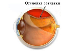
Retinal detachment
This pathology of the retina is due to retina breaks. The delaminated part of the photosensitive membrane ceases to receive power, which leads to disruption of the photoreceptors. Liquid pockets accumulate in the pockets that form, causing vision loss and continuation of retina detachment.
The detachment of the retina is:
- Regathogenic (rupture and detachment against the background of thinning of the retina);
- Tractional (against the background of the tension of the retina on the side of the vitreous body during the formation of new vessels or fibrous tissue);
- Exudative (occurs against the background of infectious diseases of the visual analyzer, neoplasms in the vascular or reticular membranes);
- Traumatic (retina can detach immediately after injury or after several months or even years after an eye injury).
Symptoms of onset of detachment:
- In one part of the field of view, a veil or shadow is formed;
- Before the eyes appear black dots;
- There are bright sparks, flashes and lightning.
Retinal detachment is treated by:
- Laser therapy (effective only in case of a break). For the prevention of detachment sometimes produce a procedure of laser strengthening of the retina;
- Vitrectomy (endoscopic surgery, accompanied by instrumental penetration inside the eye);
- Extrascleral surgery (surgery on the sclera surface).
Possible consequences: deterioration or loss of vision. Restoration of visual ability is more effective when seeking medical help immediately after the onset of symptoms of retina detachment.
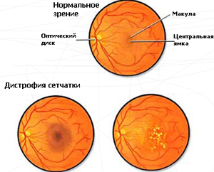
Dystrophy of the retina
Dystrophy of the retina is a degenerative, irreversible process occurring in the membrane. The disease progresses slowly, but leads to deterioration of vision, but loss of vision is rare. Pathologies are more susceptible to the elderly, in whom dystrophy is one of the common causes of impaired visual ability.
Attention! The risk group includes people with clear white skin and blue eyes. And women are more likely to encounter a problem than men.
Types of dystrophy:
- Central (affected median part of the retina, disturbed central vision);
- Peripheral (changes affect only the peripheral parts of the shell, only lateral vision suffers).
Dystrophies may be congenital or acquired. Often they are inherited from mother to child (dotted-white or twilight dystrophy, in which retina sticks are affected). The development of pathology contribute to systemic diseases of the body, as well as diseases of the visual analyzer.
Signs of peripheral dystrophy of the retina in the early stages are absent. And in the later there is a rupture of the retina, accompanied by flashes of light and swimming flies before his eyes.
With the defeat of the central zone of the retina, some areas fall out of the field of view, as well as image distortion. Symptoms may occur:
- visual disturbances in the dark;
- changes in color perception;
- blurred and blurred vision.
Treatment methods:
- Laser coagulation;
- The introduction of drugs that stop the degeneration;
- Vasoreconstructive surgery to restore nutrition of the retina through the blood vessels;
- Physiotherapy (low efficacy).
The progress of retinal dystrophy can be stopped, but vision cannot be restored after it has been disturbed due to degenerative processes.
Attention! In 2017, the first implantation of an artificial retina to a human is planned. Prior to this, the photosensitive prosthesis was tested on animals and gave excellent results. It is believed that the use of artificial retina will return sight to millions of people.
Best disease
This is the name of the degenerative process of the yellow spot of the retina. The disease occurs in children aged 5-15 years. It affects the macular area of the retina and leads to deterioration of the central vision.
Children with Best's disease do not observe any symptoms at first. But sometimes they start complaining about:
- The impossibility of reading the text printed in small print;
- Blurred vision;
- Distortion of shapes and sizes of objects in the image.
Since Best's disease is rarely accompanied by complaints from patients, it is not treated. However, such consequences as retinal hemorrhage and formation of a subretinal membrane are possible. In this case, laser coagulation is indicated.
Thrombosis of the central vein
The most important vessel that drains blood from the retina is the central vein of the retina. But sometimes occlusion, or thrombosis of this vein develops. The risk group includes people:
- Middle and old age;
- With vascular atherosclerosis, diabetes, or hypertension;
- Suffering from severe infections of the teeth or nasal sinuses.
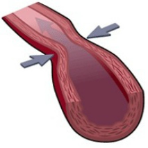
Stages of thrombosis:
- Pretrombosis. The blood flow in the vessel slows down, but the vein is not damaged yet.
- Starting thrombosis. There is a violation of blood flow in the central vein, manifested by swelling of the internal tissues of the vessel.
- Full thrombosis. The optic nerve atrophies, the retina ceases to receive nutrition.
At the first stage of thrombosis, the patient does not notice any symptoms, they will be visible only to the ophthalmologist when examining the fundus. In the second stage, retinal hemorrhage is possible. And if a patient has a damaged vein, the patient notes a decrease in visual ability.
Thrombosis of the central retinal vein is amenable to drug treatment:
- Fibrinolytics are prescribed to restore normal blood circulation in the retina (administered as an injection);
- Hormonal drugs are used topically to reduce puffiness and relieve inflammation;
- If the cause of thrombosis is hypertension, then the patient is prescribed antihypertensive drugs;
- For the prevention of re-thrombosis, antiplatelet agents are prescribed to thin the blood and reduce clotting.
Thrombosis of the central vein is dangerous with consequences in the form of glaucoma, vitreous hemorrhage, optic nerve atrophy and macular degeneration.
Retinal burn

The main cause of a retinal burn is exposure to large amounts of ultraviolet radiation. This happens when exposed to bright sunlight on unprotected eyes, or just when the light reflected from snow or water comes in. Rarely burns of the retina associated with exposure to the laser. And very rarely they appear on the background of contact with sulfuric or acetic acid in case of injury in professional conditions.
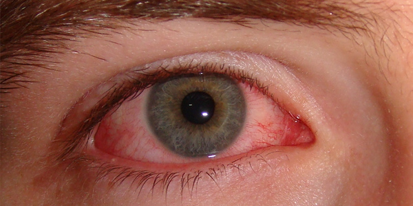
Signs of reticular burns:
- Severe redness of the eyes;
- Cutting pain in the eyes;
- Blurred vision;
- The appearance of yellow spots;
- Headache;
- Tearing;
- Swelling of the eyelids.
Rarely, only retina suffers from retinal burns. Usually it is accompanied by the defeat of many adjacent tissues. First aid for this is washing (do not use water with a chemical burn!). If the lesion is associated with exposure to bright light, then a cold compress, darkening and the use of painkillers are necessary. Restoration of the retina is possible without deterioration, and especially loss of vision.
Angiospasm of blood vessels
Retinal angiospasm is characterized by a narrowing of the lumen of the central artery of the retina or its branches. Organic changes in the blood vessels are not observed. As a result of angiospasm, retinal blood flow is temporarily limited, and sometimes it cannot get to it at all.
Angiospasm of retinal vessels is more susceptible to people suffering from:
- Raynaud's disease;
- Hypertension;
- Eclampsia;
- Diabetes mellitus;
- Atherosclerosis.
It is impossible to call angiospasm of the artery of the retina an independent disease. However, it can lead to serious consequences: blurred vision due to insufficient nutrition of the retina. With the progression of spasm may develop complete obstruction of the central artery.
Symptoms of angiospasm:
- Foggy vision;
- In the field of view appear flies;
- Violations of color perception.
Treatment of angiospasm of the central artery of the retina is the use of vasodilators, as well as drugs with a sedative and dehydrating effect.
Retinoblastoma

This name is a cancer of the retina. With this diagnosis, 1 in 20,000 children is born. The disease affects one or both (in 20-30% of cases) of the eyes and is diagnosed in early childhood. Retinoblastoma is usually hereditary, but a third of cases are associated with intrauterine eye damage due to exposure to genetically modified products or poor environmental conditions.
Retinal cancer proceeds in four stages:
- Rest Little patient does not bother. However, when examining the eye can be noted leucocoria - the detection of white pupillary reflex. This is due to the fact that a tumor appears through the pupil. Very rarely is it at this stage that a loss of peripheral or central vision occurs, and squint is more often manifested.
- Glaucoma. The child has a fear of light and an increased discharge of tears. The vessels are too filled with blood, causing the eyes to become red. The eye membranes become inflamed.
- Germination. The eyes begin to bulge due to germination of the cancer in the paranasal sinuses and the space between the membranes of the brain.
- Metastases. Cancer metastases the brain, liver, bone tissue. The patient suffers from intoxication, severe headache and persistent weakness.
Retinoblastoma is treated by:
- Drug chemotherapy;
- Radiation therapy;
- Cryotherapy;
- Laser coagulation;
- Thermotherapy;
- Conducting operations.
The prognosis for treatment of retinal cancer is favorable when pathology is detected in the first two stages. With the emergence of neoplasm and metastasis, the prognosis is poor.
Retinitis
Retinitis is the inflammation of the retina caused by an infection of the eye. The causative agents are either viruses or bacteria. Sometimes the choroidal vessels feeding the membranes of the eye are involved in the inflammatory process. Then the disease is called chorioretinitis, or retinochoryditis. The disease leads to the death of the tissues of the retina, the development of lymphocytic infiltration and the formation of scars on the membrane.
Signs of retinitis:
- Impairment of visual ability;
- Change in color perception;
- Loss of individual zones from the field of view;
- Twilight vision disorders;
- The image of objects becomes blurred and distorted;
- Flashes and lightning appear in the eyes;
- Hemorrhage occurs in the eye.
As a result of retinitis, the optic nerve may be atrophied or the retina may detach. Inflammation can be cured, but vision cannot be restored.
Treatment of retinitis depends on the cause of the disease. Drug therapy is usually performed: corticosteroids and antibacterial drugs are prescribed to the patient. In the case of a viral infection, antiviral drugs are effective. In complex therapy, vasodilators and antispasmodics are prescribed, as well as vitamins to improve blood circulation in the visual analyzer.

The retina is one of the most important structural elements of the visual analyzer. But the pathologies of the retina often lead to irreversible visual impairment, so it is important to promptly contact the ophthalmologist at the first signs of retinal diseases. In this case, the effectiveness of treatment will be the highest, and the risk of irreversible effects - minimal.
It is the retina of the eye that is most important in the structure of the human eye and has a very complex structure that ensures the perception of light pulses. She is responsible for the interaction of the visual divisions located in the brain and the optical system of the eye by transmitting and receiving visual information.
Such a disease as retinal dystrophy is caused by a disorder in the vascular system of the eye. They are sick in most cases, the elderly. The disease affects the cells of the retina - photoreceptors, which are responsible for long-range vision, as well as color perception.
The cunning of the disease lies in its asymptomatic course for some time. Sometimes the patient does not even notice his illness.
Types of retinal dystrophy
The disease can be divided into:
- hereditary or congenital;
- acquired.
Hereditary retinal dystrophy
- pigmentary - the disease is associated with a malfunction of the photoreceptors, which are responsible for gloomy vision. It is quite rare;
- point-white - occurs in early childhood and progresses with age;
Acquired retinal dystrophy
This subtype of the disease is called "senile" dystrophy or age. The development of the disease falls on the sixth dozen of a person’s life and in most cases is combined with a cataract, which is caused by peculiarities in the structure of the organism or, rather, its restructuring.
Among this disease, there are two more groups:
a) Peripheral dystrophy - develops on the background of myopia or myopia, or as a result of an injury to the eye. Due to a decrease in blood circulation in the eye, there is a decrease in the level of oxygen delivery, and with it the nutrients to the retina. It is this state of affairs that leads to the most diverse peripheral dystrophies. The disease in this form does not affect the central or macular region of the retina, which leads to an “invisible” disease, which is noticed only after the appearance of the so-called “fly” before the eyes;
b) Central Dystrophy - the disease is a flowing changes that occur in the macular region, which is the site of the most clear visual perception of objects. These include age-related macular dystrophy and serous central retinopathy. Speaking of central dystrophy, two of its subspecies are distinguished:
- Dry - provoked by metabolic products accumulating between the retina of the eye and its choroid in the form of yellow-white granules. The disease affects the cell layer, which is located under the retina, namely the retinal pigment epithelial cells. Some doctors suggest that 10 or 20 percent of patients suffering from the dry form of central dystrophy in old age will be disposed to the wet form;
- Wet - manifested through the formation of low-quality new blood vessels, through the walls of which leaks blood or intraocular fluid, the process is called "sweating." This form of the disease dramatically reduces the quality of vision, reducing its sharpness. Because of this, a huge amount of cholesterol and lipids accumulate under the retina, which provoke the "fall" of view. The form is rapidly progressive. In the end, exudative forms form subretinal scars irreversibly destroy the retina, which leads to complete loss of vision.
Causes of retinal dystrophy
Among the causes of the disease is a violation of the vascular system of the eye, which leads to the beginning of the process of scarring in the central part of the retina. As mentioned above, this disease is age-related and affects people who exchanged the sixth decade of life.
However, pathological changes are also observed in patients:
- with disturbed diet;
- tobacco abusers;
- alcohol abusers;
- suffering from changes in immune status.
Dystrophy of the retina is manifested by the lack of central vision in the patient, namely by observing objects located in the region of peripheral vision, and a black spot is observed in the center. However, lateral objects are not clearly perceived. In this position, the patient can still distinguish between day and night.
Despite the fact that retinal detachment and retinal dystrophy are somewhat similar to each other, they still represent completely different concepts and cannot be identified.
Symptoms of retinal dystrophy
Symptoms of the disease is a failure of the color perception system and in central vision. Summing up and grouping the above, it is worth noting that retinal dystrophy is accompanied by:
- reduced visual acuity;
- distortion of the perceived object;
- dark spots before eyes;
- blurred perception of the outlines of objects by the affected eye;
- violation of color perception by the sore eye;
Diagnosis of retinal dystrophy
Diagnose a disease by:
- visiometry;
- perimetry;
- eye fundus research;
- fluorescent angiography;
- eye ultrasound;
- electrophysiological studies to determine the state of the optic nerve and nerve cells of the retina;
- laboratory tests.
Risk area
While talking about such a disease as retinal dystrophy, one cannot but mention people who are at risk of this disease.
So, more than others are predisposed to it:
- Older than 50 years (women suffer more often than men);
- Falling under the hereditary factor;
- Suffering from vascular disease;
- Who do not follow the proper nutrition;
- Having problems with cholesterol;
- Tobacco abusers;
- Suffering from obesity;
- Suffering from frequent stress;
- Eating food in which there are not enough vitamins;
- Receive frequent sunburns of the eyes;
- Living in areas with problematic ecology.
Dystrophic diseases of the retina
Dystrophic diseases consist in the gradual death of retinal cells, which leads to the same gradual decrease in visual acuity. In most cases, they signal a metabolic disorder, endocrine diseases or mitochondrial.
Among the dystrophic diseases of the retina are:
- age-related macular degeneration;
- central serous choriopathy;
- diabetic retinopathy;
- hereditary dystrophy;
- pigment dystrophy;
Treatment of retinal dystrophy
To date, laser remains the most popular method of treatment. Among its greatest advantages:
- Preventing the need to open the eyeball;
- Exclusion of any infection;
- Bloodless intervention;
- Elimination of a stressful situation;
- Contactless method of exposure.
Regarding our topic will be treated with laser
- macular degeneration;
- peripheral degeneration;
- diabetic retinopathy.
Macular Degeneration
Depending on its form, which, as we have said, may be dry or wet, a treatment method is chosen:
- laser;
- surgical
With the definition of the method will help comprehensive diagnosis of the patient.
Diabetic retinopathy
It is a complication, one of the most difficult in ophthalmology, affecting visual acuity, which is observed in patients suffering from diabetes. May result in complete loss of vision due to progressive retinal damage. Lost vision can not be restored. Enough treacherous disease. It can not be seen during the passage of ophthalmoscopy or in other words, examination of the fundus. The first complaints of the patient say that the disease has gone very far, and the time of effective treatment has been irretrievably lost. Since the process of treatment is very difficult in this conversation it is impossible to describe almost nothing. A person with diabetes should be examined by an ophthalmologist once a year.
Peripheral degeneration
In case of detection of the disease, it is necessary to immediately conduct PPLC or peripheral prophylactic laser coagulation. With its help, healthy eye tissue is separated from those areas that are affected by dystrophy. In this case, a new line of attachment to the fundus of the retina is formed, and the risk of its further detachment decreases accordingly.
Treatment of retinal dystrophy folk remedies
In this section, we present the most common treatments in traditional medicine.
Leeches
The treatment consists in the properties of their saliva, which is injected into the blood during skin penetration. It contains a huge amount of enzymes and has a whole range of effects.
Leech secret has the following actions:
- anti-inflammatory;
- painkillers;
- improves the immune system;
- pressure reducing;
- lowers blood sugar and cholesterol levels;
- detoxifying;
- improves microcirculation.
Goat milk
Mix it in the same proportion with water. Bury one drop in each eye. After that, wear light-transmitting fabric over your eyes for 30 minutes. Treatment continue for seven days. The procedure is designed to prevent retinal detachment.
Broth from the berries of wild rose, onion peel and needles
All the above ingredients are crushed and mixed in a ratio of 2: 2: 5. The mixture is poured with one liter of water and boiled for 10 minutes. The broth is taken in the amount of 0.5 liters per day for thirty days.
Cumin broth
A tablespoon pour 200 ml. boiling water and heated for five minutes on low heat. After that, add 1 teaspoon of cornflower to the broth and mix. Leave to cool. Bury the broth eyes, two drops, during the day twice.
Infusion of mustard, horsetail, cranberries and birch leaves
Celandine
It is a great healing plant. One teaspoon of celandine is crushed and poured 100 ml. water. The contents are held for a few seconds on the fire and then allowed to infuse. Filter and place the contents in the refrigerator. Bury their eyes three times a day, three drops for one month.
The reticular membrane of the eyeball is the layer responsible for the visual perception of the environment. Diseases of the retina have serious consequences that affect visual acuity. In the advanced stage, without proper medical treatment, ninety percent of cases lead to complete blindness.
A certain age group of risk in diseases of the eyeball does not exist, it affects both the elderly and newborns. The risk of developing a disease associated with the eye membrane, there is in people suffering from myopia and diabetes. Diagnosis of the disease at an early stage allows for timely medical intervention and to stop the development of pathology.
Changes affecting the structure of the eyeball have different causes. In some situations, it is not possible to identify retinal diseases in the early stages.
The disease affects the following areas:
- The central part - here are located: the vascular system and the optic nerve.
- The peripheral part is the region of photoreceptors consisting of rods and cones.
Causes of the disease
Damage to the retina can be caused by various injuries, the occurrence of inflammatory processes, infections and myopathy. The presence of the following diseases may cause the start of pathological changes:
- hypertension;
- diabetes;
- atherosclerosis.
For example, retinopathy - developed against the background of diabetes mellitus, a disease that is not amenable to treatment. The progression of the disease can only be blocked, however, it is not possible to completely restore vision.
The first symptoms of a disease of the retina can manifest themselves in the form of various “flies” in front of the eyes, loss of color perception and sharpness of vision. However, in many cases in the initial stages of the disease does not manifest itself.
Dystrophy of the retina of the eyeball
A characteristic feature of the disease is a violation of the vascular system of the eyeball. This retinal disease in older people is diagnosed quite often. In addition, a high degree of myopia can also lead to the development of the disease, as the visual organs increase in size. In the wake of an enlarged eye, the retina increases, stretching and thinning. Symptoms may be the following:
- loss of sharpness of perception;
- problems with visual perception at twilight;
- problems associated with the disappearance of peripheral vision.
In order to stop the progression of the disease, timely intervention is necessary. Usually, the dystrophy of the reticular area of the eyeball is treated with laser equipment. The retina is soldered to the vessels with the help of a laser in order to avoid its rupture. The operation is harmless and does not have disastrous consequences for the body.
 All diseases can be divided into three groups: dystrophic, inflammatory and vascular
 Retinal tumor
The disease is divided into two categories:
- benign;
- malignant.
The disease is hereditary and in seventy percent of cases manifests itself in the age of one year. Often the disease affects both visual organs. The first stages of development show no symptoms, and are detected only during the ultrasound procedure. Without the necessary treatment, the tumor is localized throughout the intraocular area.
For an absolute victory, it is necessary to start treatment immediately after its diagnosis. For treatment, the method of freezing and photocoagulation is used.
Hemorrhage
Damage to the vascular system can lead to such consequences as vision loss, retina dystrophy, retinal detachment, and glaucoma formation. The cause lies in the problems associated with blood vessels and clogged arteries.
This type of disease can be caused by the effects of diabetes mellitus, heart muscle work problems, as well as mechanical injuries and injuries. Patients complain of deterioration of perception, and sensations of spots in the eyeball. Treatment can be carried out both with the help of medicines and surgery.
Damage to the vascular system
This category includes most of the diseases that provoke changes in the structure of the vascular system. Damage to the blood vessels is in the first place in the list of diseases that lead to total blindness. Diseases lead to impaired metabolism of the eyeball's nutrients, which leads to disruption of the photoreceptors. The disease is dangerous by the development of various types of blood clots.
 Dystrophic diseases of the retina are the most common.
 Peripheral degeneration of the retina
This violation of the retina leads to the appearance of thinned areas, as a result of which gaps appear. Complicated stage of the disease can lead to detachment of the reticular layer and loss of visual perception. Modern medicine is fully capable of influencing the disease process and stopping the devastating effects.
People at risk of suffering from myopia are at risk. Increasing the size of the eyeball, slows down the flow of blood into the blood vessels, and the retina, stops receiving the required amount of nutrients. Its structure becomes loose and heterogeneous. The appearance of various outbreaks in the eye can be a precursor of the disease.
The disease is genetic. Constant nervous stress, poor ecology, infections, physical exertion, complications during pregnancy - all this can lead to the appearance of the first symptoms. Symptoms of retinal disease, a frequent occurrence for the elderly. That is why it is very important to regularly diagnose the disease at the ophthalmologist.
Retinal disinsertion
Retinal detachment of the eyeball is a pathology that requires immediate surgical treatment. The disease consists in the discharge of the retina from the sheath consisting of vessels. The result may be a violation of the entire blood supply to the organs of vision and the death of photoreceptors. Without timely surgical intervention, always causes complete blindness.
The outer parts of the retina have a tight connection with the vitreous body. Natural aging of the body leads to a decrease in the size of the vitreous body. When it is disconnected from the mesh region, a gap appears, into which the fluid penetrates. The disease can be caused by:
- the result of traumatic brain injury;
- mechanical damage to the eyeball;
- consequence of surgical intervention;
- dystrophy of the optic organ;
- myopia.
 Damage to the inner lining of the eye due to various factors.
 Retina tear
To tears of the retina, most often prone to people suffering from myopia, as the development of the disease affects the entire structure of the eyeball. Symptoms of the disease are bright flashes of light in the eye and the appearance of black threads. At an early stage, the edges in the area of the gap begin to peel off, and at later stages, the retina peels off completely.
For treatment in the early stages of the disease laser technique is used. The affected areas are strengthened by laser coagulation. In the areas subjected to such an impact, “adhesions” are formed, the purpose of which is to create a strong connection between the reticular region of the eyeball and the vascular system.
Macular Degeneration
Macula - a segment of the eye, having the form of a sphere. Here is a huge number of receptors. Macula plays a large role in the visual processes that occur when a person focuses his or her vision on closely spaced objects. Macular degeneration is the process of the formation of pathology, which leads to a strong decrease in the quality of perception. The initial stages of the disease are accompanied by the following symptoms:
- curvature of the shape of objects;
- appearance of veil in the visual area;
- difficulty in reading, due to the loss of letters;
- dimming perception.
The disease is divided into two forms: dry and wet. The dry form of the disease is accompanied by the slow development of degenerative changes that originate from problems with blood circulation. The wet form of macular degeneration primarily affects the vascular system. The body begins to create defective vessels, the walls of which are very thin. Through such vessels, fluid enters the retina, which causes swelling and hemorrhage. At a later stage, the appearance of microscopic scars is possible, which disrupts the work of the central vision.
 The main symptom of retinal disease in humans is the so-called veil
 Treatment of macular degeneration directly depends on the stage of the disease at which the patient is seeking help. Depending on this factor, the ophthalmologist may choose the form of exposure.
The laser effect used in the treatment of retinal diseases was discussed above. Another method may be the introduction of drugs directly into the vitreous body by injection. The drug has blocking properties and does not allow developing affected vessels to grow. Today, most medical centers use drugs such as LUCENTIS and EILEA.
Retinitis
Retinitis is an inflammation of the retina, which can be both unilateral and bilateral. The disease is infectious or allergic. The cause of the disease can be syphilis, the presence of purulent and viral infections, AIDS.
Depending on the location on the retina, the disease may exhibit different symptoms. The main ones that can be summarized are the gradual deterioration of the quality of vision and the narrowing of the visual field. In a small proportion of cases, the disease is localized in small areas, subsequently spreading throughout the retina. Late diagnosis of the disease can cause vision loss. Experts recommend treating retinitis with a number of medications.
 All diseases of the retina develop painlessly, since the inner lining of the eye does not have sensitive innervation.
 Angiopathy
Angiopathy of the eyeball - a disease that affects the vascular system, most often the disease is a consequence of dystonia, hypertension and diabetes mellitus.
Damage to the circulatory system is most often expressed in spasms and cuts in the visual organs.
The effect of diabetes
Diabetic retinopathy is a disease caused by diabetes. During the course of the disease, the vascular system of the eye retina is affected. The first symptoms may be:
- the appearance of floating spots;
- the appearance of veil;
- mist before eyes
The late stage of the disease is characterized by complete loss of vision. Long-term development of diabetes leads to damage to the vascular system of the eyeball. The vessels become thinner, many capillaries are clogged, and the newly appeared vessels have a damaged structure. Very often scars appear in the area of the affected tissues. According to the research it was found that people suffering from diabetes for many years, one hundred percent susceptible to retinopathy.
Conclusion
The presented list is only a small part of the diseases associated with the organs of sight. Problems with the retina of the eyeball can have the character of burns, edema and traumas of the retina, which in most cases has a detrimental effect on the visual organs. Treatment of diseases of the retina, it is very important to carry out in a timely manner, and only in this case you can count on a positive result. In order to keep your eyesight healthy, you need to visit the office of an ophthalmologist at least once a year.
- In contact with 0
- Google+ 0
- OK 0
- Facebook 0