Color vision abnormalities
Anomalies are usually called those or other minor violations of color perception. They are inherited as a recessive trait linked to the X chromosome. Persons with a color anomaly are all trichromates, i.e. they, like people with normal color vision, for a full description of the visible color, you must use the three primary colors. However, the anomalies are worse distinguished by some colors than trichromates with normal vision, and in the color matching tests they use red and green in other proportions. Testing on the anomaloskop shows that when the protonomaly in the color mixture is more red than normal, and when deuteranomaly in the mixture more than you need, green. In rare cases of tritanomalia, the yellow-blue channel is impaired.
Dichromates
Various forms of dichromatopsia are also inherited as recessive X-linked traits. Dichromates can describe all the colors they see with just two pure colors. Both the protanopes and the deuteranopes have a disrupted red-green channel. The protanopes confuse red with black, dark gray, brown, and in some cases, like deuterorans, with green. A certain part of the spectrum seems to them achromatic. For protanop, this region is between 480 and 495 nm, for deuteranop - between 495 and 500 nm. Rarely encountered tritanopas confuse yellow and blue. The blue-violet end of the spectrum seems to them achromatic - like a transition from gray to black. The spectral region between 565 and 575 nm is also perceived by tritanops as achromatic.
Full color blindness
Less than 0.01% of all people suffer from complete color blindness. These monochromats see the world as a black and white film, i.e. distinguish only grayscale. Such monochromats usually have a violation of light adaptation at the photopic level of illumination. Due to the fact that monochromatic eyes are easily blinded, they poorly distinguish the shape in daylight, which causes photophobia. Therefore, they wear dark sunglasses even with normal daylight. Histological examination usually does not find any anomalies in the retina of monochromats. It is believed that their cones instead of visual pigment contains rhodopsin.
Violations of the stick
People with anomalies of the kidney apparatus perceive color normally, but their ability to adapt darkly is significantly reduced. The reason for such “night blindness”, or niktalopii, may be insufficient content in the consumed food vitamin A1, which is the starting material for the synthesis of retinal.
Diagnosis of color vision disorders
Since color vision disorders are inherited as a sign linked to the X chromosome, they are much more common in men than in women. The frequency of protanomaly in men is about 0.9%, protanopia - 1.1%, deuteranomalia 3-4% and deuteranopia - 1.5%. Tritanomalia and tritanopia are extremely rare. In women, deuteranomalia occurs with a frequency of 0.3%, and protanomaly - 0.5%.
Normal picture:

Deuteranope (lack of red-green):


Protanope (another form of red-green deficiency):


Tritanope (lack of blue and yellow, very rare form):
![]()

keep in mind that this shows the ULTIMATE options (well, if there is no sensitivity for these colors at all)
Want to test yourself?
There are tables of Ishihara, for eye examinations, selected from random circles so that dichromates (two-color vision) and trichromats (three-color, high-grade) and non-chromats see different numbers / pictures on these test tables.

Figure 1. All normal trichromates, anomalous trichromates and dichromates distinguish the numbers 9 and 6 (96) equally correctly in the table. The table is intended primarily for demonstration of the method and for control purposes.
![]()
Figure 2. All normal trichromats, anomalous trichromates and dichromates distinguish two figures in the table equally correctly: a triangle and a circle. Like the first table, it is intended primarily for demonstration of the method and for control purposes.

Figure 3. Normal trichromats distinguish the number in the table 9. Protanopes and deuteranopes distinguish the number 5.

Figure 4. Normal trichromats distinguish a triangle in the table. Protanopes and deuteranopas see a circle.

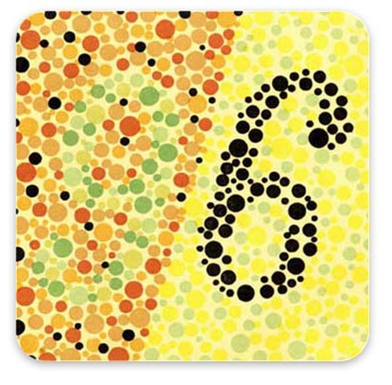
Figure 5. Normal trichromates distinguish figures 1 and 3 in the table (13). Protanopes and deuteranopes read this figure as 6.

Figure 6. Normal trichromats distinguish two figures in the table: a circle and a triangle. The protanopes and deuterorans do not distinguish these figures.

Figure 7. Normal trichromats and protanopes distinguish two numbers in the table - 9 and 6. Deuteranopes distinguish only figure 6.

Figure 8. Normal trichromats distinguish the number in the table 5. Protanopes and deuteranopes distinguish this figure with difficulty, or do not distinguish it at all.

Figure 9. Normal trichromats and deyraneopes distinguish the number in the table 9. The protanopes read it as 6 or 8.

Figure 10. Normal trichromates distinguish figures 1, 3, and 6 (136) in the table. The protanopes and deuteranopes read two numbers 66, 68, or 69 instead.

Figure 11. Normal trichromats distinguish a circle and a triangle in the table. The protanopes distinguish a triangle in the table, and the deuteranopes distinguish a circle, or a circle and a triangle.

Figure 12. Normal trichromats and deyraneopes distinguish figures 1 and 2 in the table (12). Protanopes do not distinguish these numbers.

Figure 13. Normal trichromats read a circle and a triangle in the table. The protanopes distinguish only the circle, and the deuteranopes, the triangle.

Figure 14. Normal trichromats distinguish the numbers 3 and 0 (30) in the upper part of the table, and in the lower part they do not distinguish anything. The protanopes read the numbers 1 and 0 (10) in the upper part of the table, and the hidden number 6 in the lower part. The deytranopes distinguish the number 1 in the upper part of the table, and the hidden figure 6 in the lower part.

Figure 15. Normal trichromats distinguish two figures at the top of the table: a circle on the left and a triangle on the right. The protanopes distinguish two triangles in the upper part of the table and a square in the lower part, and a deuteropenop in the upper left triangle, and in the lower part a square.
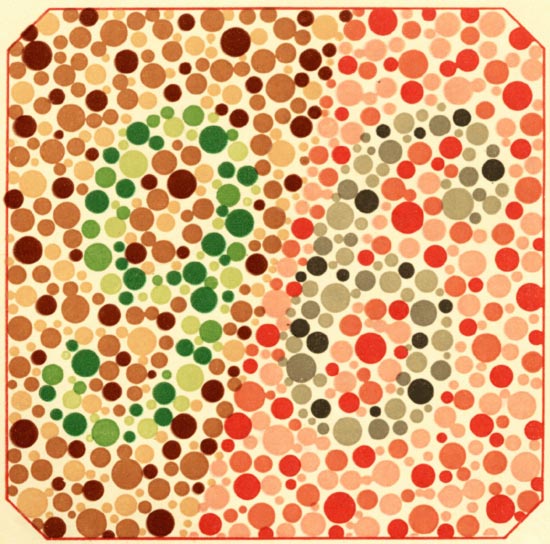
Figure 16. Normal trichromats distinguish in the table the numbers 9 and 6 (96). The protanopes distinguish in it only one digit 9, deuteranopes - only digit 6.

Figure 17. Normal trichromats distinguish two shapes: a triangle and a circle. The protanopes distinguish the triangle in the table, and the deuteranopes - the circle.

Figure 18. Normal trichromats perceive horizontal rows in the table of eight squares in each (color rows of the 9th, 10th, 11th, 12th, 13th, 14th, 15th and 16th) as monochromatic ; vertical rows are perceived by them as multicolored. Dichromats, on the other hand, perceive vertical rows as monochromatic, with protanopes accepting as monochromatic vertical color rows — the 3rd, 5th, and 7th, and deuteranopes — vertical color rows — the 1st, 2nd, 4th, and 6th. th and 8th. The colored squares located horizontally are perceived by the protanopes and deutero-pami as multicolored.
Color blindness or color blindness is a violation of color vision. With this pathology, the eye is not able to distinguish one or more shades or primary colors.
Most often it is hereditary, associated with disorders in the X chromosome. Color blindness is transmitted through the female line from the mother, who is a carrier, but it is mainly men who suffer from it.
In some cases, this disease is acquired. Caused by diseases associated with the optic nerve or retina and is not inherited. The patient has difficulty distinguishing between blue and yellow. Cataracts are the most common cause of acquired color blindness.
In the retina there are special nerve photosensitive cells - cones. They provide adequate color perception. There are 3 types of cones. Each of them is responsible for the perception of one of the primary colors: blue, green, red.
In normal color perception, all 3 types of receptors are involved. Such people are called trichromats. In most cases, color blindness does not distinguish between one or two of the primary colors. If a person does not distinguish any color - this is color blindness.
If a person does not distinguish between red color, this is protanopia Does not distinguish between blue color - tritanopiya (extremely rare). Does not distinguish between green color - deuteranopy.
The only symptom of color blindness is a violation of color perception. Severe forms, such as the complete absence of color vision, are not common. Often the patient is not able to distinguish only shades.
Despite the fact that the symptoms of this disease are already apparent in childhood, a person may not notice any deviations in color perception. Occasionally, it is necessary to examine the oculist in order to detect this violation.
The color blindness test is a collection of polychromatic tables. Each of them shows the same in color, but slightly different in color, colored circles.
The picture will appear homogeneous to a person who does not distinguish the colors present on it. A person with normal vision will see numbers or geometric figures, in which circles of the same color are combined.
Test
- To check the color vision, move 1 meter away from the computer.
- Relax, look at each picture for no more than 5 seconds.
- If you get a negative result, do not be discouraged. Perhaps the reason is the color of your computer monitor. However, it would not be superfluous to consult a specialist.

A person with normal vision (trichromate) sees the number 15 in the picture. A person who does not distinguish between green (protanop) or red (deuteranop) sees the number 17. If there is nothing visible in the picture, this is complete color blindness.

A person with normal vision sees the number 26 in the picture. A person with impaired perception of the red part of the spectrum (protanopia) sees the number 6 and, possibly, 2. A person with impaired perception of the green part of the spectrum (deuteranopia) sees 2 and possibly 6.

People with the usual color perception do not see anything in this picture. When a violation of the perception of red or green color, a person sees the number 45

A person with ordinary vision can distinguish an image of a continuous chain in this picture. A person with disturbances in the perception of green or red color cannot distinguish a continuous chain.
A person with a normal perception of color sees the number 29 in the picture. A person who does not distinguish between green or red color sees the number 70.

The figure shows the number 5. It can be distinguished as a person with normal perception, and with blindness in the red or green parts of the spectrum. But people with impaired color perception will be able to see it with difficulty or not see it at all.

The figure shows the number 73. It is quite able to distinguish both people with normal perception, and with blindness in the green or red parts of the spectrum. But people with impaired color perception will be able to see it with difficulty or will not see it at all.

A person with a normal color perception sees the figure 8 in the picture. If blindness is present in the red or green parts of the spectrum, the person sees the number 3.
Eyesight test for color perception is obligatory for people of some professions. With the help of special tests, one or another form of color blindness can be identified in the subject, which will serve as an obstacle to obtaining the specialty of a pilot, a driver of military equipment, a train driver, a seaman and some medical directions. A person whose eyes perceive color in a somewhat distorted way will not fix the signals on the road, the map or will not be able to control complex devices.
Testing is carried out by showing pictures consisting of multi-colored circles forming figures and certain figures. If there are problems with color perception (color weakness, blindness to certain colors), a person may not see the figures, distinguish not all the numbers or visually perceive completely different signs.
Important test conditions. The person should feel good, the room is needed for these purposes, illuminated, and the distance is convenient for the person being tested. Recognition time - no more than 10 seconds.
Test number 1
Absolutely all people see the numbers 9 and 6. This is the so-called checklist used to identify simulators.
Test number 2

A person with a normal color perception (trichromate) sees the figure 13 in the picture, and people who do not distinguish between the red color (protanopa) or the green color (deuteranopes) will see the number 6.
Test number 3
People with normal eyesight and those with impaired perception of red distinguish between two numbers - 9 and 6 (96), and a person with a violation of green perception will see only the number 6.
Test number 4

Normally, figures 1, 3 and 6 (136) are distinguishable in the picture. And if color perception is disturbed, only two numbers are distinguishable (for example, 69, 68 or 66).
Test number 5

Test number 6
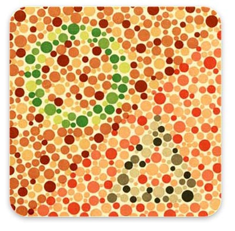
A person with normal vision distinguishes between a circle and a triangle. If the perception of red is disturbed, only the circle is visible, and if the perception of the green is disturbed, only a triangle.
Test number 7

Normally (trichromasy) there are two digits (3 and 0) at the top of the image. If the perception of red is disturbed (protoanopia), the person in the upper part of the table sees the numbers 1 and 0, and in the lower part - 1 (normally not visible). In case of violation of the perception of the green color (deuteranopia), the figure 1 is distinguishable at the top, and the figure 6 at the bottom.
As a rule, the cause of color blindness is a genetic defect of vision, which manifests itself most often in men. A pathological anomaly of color perception may occur in the process of aging of the body, as well as as a result of injury or illness.
It is important to bear in mind that this test for color perception is considered approximate and cannot categorically indicate the absence or presence of a violation of color vision. If you need a more accurate diagnosis, consult an ophthalmologist.
If the material seems useful to you, please share him with friends.
- In contact with 0
- Google+ 0
- OK 0
- Facebook 0