Eye hemorrhage - It is a collection of blood that has poured from the damaged vessels into the surrounding tissues. It can be caused by injury to the eye or head, diseases associated with impaired blood circulation or damage to the walls of blood vessels, excessive physical exertion, or other causes.
In order to understand what to do and how to treat a hemorrhage in the eye, one must first determine in which structure of the eye this has happened. Symptoms of hemorrhage in the eye vary depending on the localization of the pathological process.
Retinal hemorrhage
The main symptoms of retinal hemorrhage are:
- reduction;
- image blur;
- or “mesh” before the eyes;
- headache in the temple with the affected eye.
Visible manifestations in this type of ocular hemorrhage may be absent. If the hemorrhage is single and not extensive, rest is recommended as a treatment for the eyes, hemostatic and vasoakreplyayuschie drugs are prescribed. In severe cases - with hemorrhage, which occupies a large area and is often repeated, hospitalization is required in the ophthalmology department. Recurrent retinal hemorrhages can lead to blindness.
Hemorrhage in the sclera (white) eyes
The following symptoms indicate the accumulation of blood in the protein shell of the eye:
- pain in the eye;
- slight decrease in vision;
- red spot on the surface of the sclera.
In this case, special treatment is not required, the accumulation of blood resolves itself within 48 - 72 hours.
Vitreous hemorrhage
Vitreous hemorrhage is called hemophthalmus. Symptoms of this process are as follows:
- visually visible uniform formation of brown color behind the lens of the eye;
- the appearance of dark moving spots before the eyes;
- flashes of light before your eyes.
This pathological process occurs when damage to the choroid with blood entering the vitreous body. In this part of the eye there is no possibility of delimitation of the physiological fluid, therefore its rapid turbidity occurs. Full hemophthalmus can lead to loss of vision, if during the first hours after hemorrhage medical care is not provided. Severe complications are also possible - for example, retinal detachment.
Hemorrhage in the anterior chamber of the eye
Hemorrhage in the anterior chamber of the eye, or hyphema, is characterized by such signs:
- the appearance on the eye of a homogeneous red formation with smooth edges;
- reduced visual acuity.
With this type of hemorrhage in the eye, blood fills the space between the cornea and the iris. In most cases, blood resorption occurs spontaneously within a few days. To speed up this process, a resorptional treatment may be prescribed. It should be borne in mind that in case of hyphema it is necessary to exclude the use of nonsteroidal anti-inflammatory drugs and anticoagulants, since they can disrupt the coagulation system.
If hyphema does not go away after 10 days, then this may indicate the development of complications, which include:
What to do if there is a hemorrhage in the eye?
At the first sign and suspicion of hemorrhage in the eye (even a slight, at first glance), an urgent need to contact an ophthalmologist or a general practitioner. For the diagnosis of pathology, a series of studies will be carried out, which, in addition to an ophthalmologic examination, necessarily include a blood test (total and for sugar). After that, appropriate treatment is prescribed.
Why does the eye fill up with a red spot, blood pressure and other causes of this manifestation - these questions arise in patients with a characteristic ailment. The thinnest mesh of blood vessels between the sclera and conjunctiva is extremely sensitive to the slightest pressure drop and physical exertion. In these and other cases, there is a local destruction of the vascular walls, which is why the eye floods the red spot. Blood pressure can be both a cause and a consequence. Local hemorrhage in the eye is a rather unpleasant phenomenon and it can also cause and be caused by a number of dangerous diseases.
1 Types and symptoms of hemorrhage
Eye hemorrhage is of several types:
- Retina: it is characterized depending on the degree of damage, usually the contours of the observed objects are washed out, many small bright light spots - "flies" flash, bloody veil, with frequent regular repetition threatens visual loss.
- In the eye socket: a very serious damage to the eye due to a contusion of its orbit; may be associated with vasculitis and blood disorders. Symptoms of this damage may include a bug-eyed, restriction of eyeball movements or pushing it forward, eyesight impaired, at the turn of the base of the skull of the eye it is filled with blood according to the type of glasses.
- To hyphema (anterior chamber of the eye): with this hemorrhage, the blood stain has even contours, can be localized in the fundus (if the person is standing), or the eye looks as if it has been completely filled with blood (in the supine position). Not as dangerous as it may seem at first glance, clots quickly dissolve, and vision almost does not suffer.
- In hemophthalmus (vitreous body): causes a dense red spot to leak out as a result of total damage to the eye vessels right behind the lens of the eye. Very dangerous such complications as retinal detachment or atrophy of the eyeball, which usually leads to complete blindness. Such damage requires immediate medical attention and is accompanied by flashes of black dots or white spots.
2 Causes of pathology
Let us consider several main causes provoking damage to the ocular vessels and, as a result, hemorrhage:
- Mechanical external damage (contusion) of the eye or skull and even the chest: distinguish between mild, moderate and severe severity of blunt trauma.
- External stimuli: these include wind, dust, pollen, tobacco and other smoke, chemicals, cosmetics, etc., can cause irritation of the eye mucosa and rupture of small capillaries.
- Prolonged tension of the organs of vision: reading, watching TV, computer screen for quite a long period without interruption or in poor light.
- Insufficient rest of the whole body: due to insomnia, stress, fatigue, etc.
- Neglect of the rules of wearing contact lenses.
- Physical exertion and sharp spasms in the chest: the latter may include coughing, leading to conjunctival hemorrhage, as well as screaming, attempts during labor or bowel problems, uncontrollable vomiting and cardiovascular disease.
- Side effect after surgery: as a natural reaction of the body, expressed in the form of an eye, which became bruised.
- Local inflammation in the eyes.
- Neoplasms (tumors) in the eye: constantly expanding tumor tissue can squeeze the blood vessels in the eyes to the point of no return.
- Infectious eye diseases: blepharitis, conjunctivitis, uveitis.
- Pathologies of the cardiovascular system: vascular atherosclerosis, hypertension (pressure surges can lead to local rupture of small vessels), a decrease in the level of collagen protein in the blood.
- Diabetes: it develops a complication of the vascular system - retinopathy.
- Lack of vitamins: A, B2 and B6 leads to disruption of the normal functionality of the vessels, in particular the outflow of excess fluid from the blood.
- Individual features of the anatomy: too "gentle" structure of the eye vessels, thin shell of the eye, easily irritated even from unhealthy diet, sleep and rest, etc.
3 First aid and treatment of the disease
If the eye is covered with a red spot and the more partial or complete loss of vision is observed, you should immediately consult a doctor.
This is especially true for such cases in which the redness does not pass more than 2 days, pain, burning, foreign body is felt in the eye area, photophobia or yellow (green) discharge appears.
Something you can do yourself, for example, as a temporary measure to use the "artificial tear", put a cold compress on the eye area, using cold water, ice, potatoes, chamomile, cornflower or ordinary tea leaves. At the same time, the vessels will narrow slightly, and the inflammation will decrease.
It should be understood that with exclusively mechanical damage these measures are even contraindicated, especially if there is a foreign body or its fragments in the eyes. Then, before the arrival of the doctor, it is imperative to impose a recent sterile gauze bandage on the affected area in order to prevent additional infection.
Treatment of hemorrhage in the eye. First, after examining and determining the extent of damage, the doctor takes measures to stop the bleeding and eliminate blood clots. Then the root cause of hemorrhage is surely found out, usually through a general blood test.


Sometimes a person has blood in the eye, the reasons for this lie in the violation of the integrity of blood vessels.
Causes of blood
The most common causes of blood in the eyes can be called injury or contusion. That is, bleeding from the blow to the head or damage to the organ of vision may appear in the eye.
If you have even an insignificant amount of blood in your eye, you need to consult a doctor, as this can cause serious pathologies and diseases, and then you can’t do without surgical intervention.
Often, a closed brain injury can cause eye disease.
In medical practice there are three types of contusion:
- Light: when the eye remains intact, and vision is restored over time.
- Medium: when the structure of the eye is damaged, and vision is reduced to light sensations.
- Severe: this is the most serious degree of the disease, can develop up to the loss of vision.
But not only mechanical damage can provoke bloodshot eyes, it can also occur due to certain diseases of the internal organs, for example, due to disturbances in the circulatory system.
Bloodshot eyes may indicate an increase in blood pressure and atherosclerosis. If the venous artery thickens, then the elasticity of the vessels may be disrupted.
In medicine, such cases are known when blood flows from the eye due to diabetes mellitus, which causes changes in the retina of the eye: the vessel wall begins to weaken, and as a result, blood is poured into the eye.
Hemorrhage can overtake a person due to low hemoglobin and leukemia.
The following actions can provoke the appearance of blood from the eye:

- attempts at childbirth;
- strong loads on the body;
- scream;
- cough.
Blood can be released in different parts of the eye, such as:
- front camera;
- vitreous body;
- retina;
- eye socket.
Why do vessels burst
Is the eye filled with blood? This may occur due to the fact that the vessels burst. This problem is quite common and has the following symptoms: redness, itching, burning.
 The vessels in the eyes are thin, they can easily burst. This can occur due to eye strain and fatigue, so you should constantly give your eyes a rest, especially if you work at a computer for a long time.
The reasons for which vessels can burst:
- light from the lamp;
- insomnia;
- long reading in the dark;
- long stay in rooms with air conditioning.
To keep the vessels from bursting, the eyes must rest from time to time. So, if you work at a computer, you should rest your eyes every twenty minutes. In such breaks you need to relax, do not read and watch TV. You can drink tea, take a bath, just lie down or talk to someone.
It is worth as much as possible to eat vegetables, fruits, preferably green, as these products contain the most substances that help to strengthen the eyes.
Sometimes, to strengthen the blood vessels, the doctor prescribes Vizin drops, which are completely harmless to humans. If you often burst blood vessels, then you need to pass an analysis on the sugar content in the blood.
Blood in the anterior chamber
 When blood is poured into the anterior chamber, this disease is called hyphema. It can flow in different ways.
Most often, the development occurs as follows: red formations appear in the eye, differing in even contours, blood begins to occupy the entire surface of the eye.
If a person spends a lot of time in an upright position, then blood can settle on the bottom.
With such symptoms, vision is usually not impaired, the blood is absorbed after a few days. This phenomenon occurs in many people.
Blood in the Vitreous
When blood enters the vitreous, a geophthalmos occurs. This disease can only be talked about if the eye shell is damaged.
Blood in the retina
 If blood enters the retina, the development of the disease will depend only on the extent to which the retina is damaged. People with such pathologies complain of the following:
- blurred image;
- the appearance of the mesh;
- blurred vision;
- flickering before the eyes.
If hemorrhages are frequent, the patient may completely lose his sight.
The eye is swollen and at the same time blood is seen in it - then this can happen because of the contusion of the orbit, in which case it is rarely possible to avoid beoglazy. Also, with these symptoms, an exophthalmos can develop when the eyeball moves forward. Then the patient may complain of the following symptoms:
- in the eyes of two;
- limited eye movement;
- blood begins to flow under the eyelid.
 Since the time when the eye is damaged, it can take up to a day, there are also cases when more than a day passes before the appearance of circles under the eyes. If you start treatment on time, you can save your eyesight and avoid complications of various kinds.
Usually eye damage in hyphema does not require treatment, you need to better monitor the patient's condition and, at the first suspicion, you should call the doctor so that he can assess the person's condition.
Sometimes, for prevention, the doctor may prescribe iodide drops. Instill the drug three times a day, one drop in the eye. The course of treatment must last at least one week.
If the symptoms of the disease continue for more than two weeks, then you need to consult a doctor for advice.
This is especially true for older people, as they can lose their sight in seconds. Such symptoms can lead to complications of the following kind: tuveitis, glaucoma, cataracts.
If the doctor prescribes treatment, then you should refrain from the use of anti-inflammatory and non-steroidal drugs.
In that case, if the hemorrhage was only once, and the shape of the eye has not changed, and the patient feels good, then this disease will not require special treatment. All that is needed is rest and rest to the eyes. People with such diseases are sometimes prescribed vascular stiffening and hemostatic drugs.
If it is a hemorrhage in the retina, the patient requires medical care and hospitalization.
Take care of your youth and be healthy!
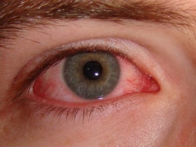
Redness of the eye is a sign of minor irritation or a dangerous infection process. There are several reasons for red eyes.
A person's eyes turn red, "poured" with blood, when small blood vessels on the surface of the eyes expand and overflow. This phenomenon is associated with an insufficient supply of oxygen to the cornea.
Bloody eyes themselves are not a reason to declare anxiety. But if the reddening of the organs of vision is accompanied by pain, visual impairment or abnormal discharge from the eyes, it indicates a serious medical problem.
The conjunctiva is the mucous membrane of the eye, a thin transparent membrane covering the eyeball. Its inflammation is called conjunctivitis, it usually develops due to infection, adenoviral or bacterial.
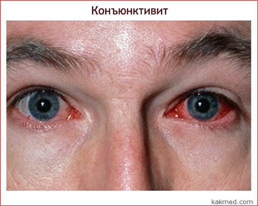
Contamination of the conjunctiva with pathogens leads to irritation of the blood vessels, causing them to swell. The white of the eye is colored because of this in a reddish and even pinkish color. Up to 80% of cases of conjunctivitis are associated with viruses. Adenovirus conjunctivitis often affects children in schools and other institutions where viruses are easily transmitted from healthy patients. Not even easy, but very.
The infection is transmitted through fingers and personal items. Often, a viral infection also causes a cold catarrh of the respiratory system, in which case the pathogen is spread by airborne droplets, like flu.
Allergic conjunctivitis occurs due to allergies, such as dust. Prolonged wearing of contact lenses or poor cleaning of them also lead to inflammation of the mucous membrane of the eyes.
If the infection got into one eye, then soon it turns out in both, because it is easy. As a result, both eyeballs turn red.
The list of conjunctivitis symptoms is as follows:
- itchy eyes
- excessive discharge of tears
- eye redness
- abnormal discharge
- blurred vision
- feeling of "sand" in one or both eyes.
Diagnosis and treatment of conjunctivitis
The doctor makes a diagnosis of conjunctivitis on the well-known pinkish shade and the nature of the discharge from the eyes, if any. With a bacterial infection of the mucous membrane of the red may not be, but there are abnormal discharge from the eyes - white, green or yellow. To confirm the diagnosis, samples of ophthalmic secretions are taken for analysis to the laboratory.
If conjunctivitis is allergic, conduct a study to establish a specific allergen, which the patient should continue to avoid.
The method depends on its nature, so it is important for a specialist to be able to make a diagnosis. Conjunctival inflammation usually resolves without treatment and without any consequences for vision. But if you have the above symptoms, you need to be alert, because some pathogens are seriously dangerous. For example, the herpes virus.
In order not to become a carrier of conjunctivitis, it is necessary to wash hands more often and try not to rub their eyes. Avoid general use of such things as eye drops, cosmetics, towels, or pillowcases.
When a bacterial or viral conjunctivitis resolves, contact lenses, solutions for them, and / or eye makeup that the patient used while being contagious are thrown away. So you can avoid re-infection.
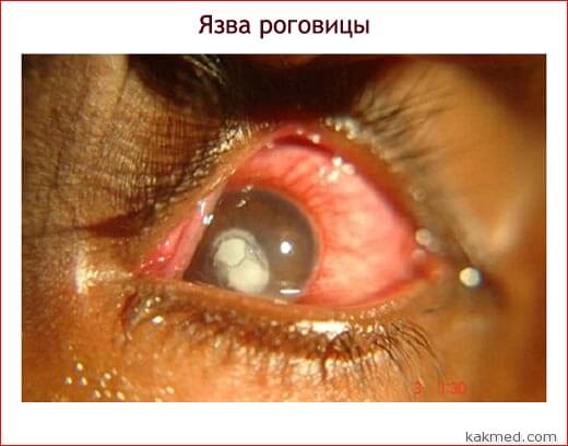
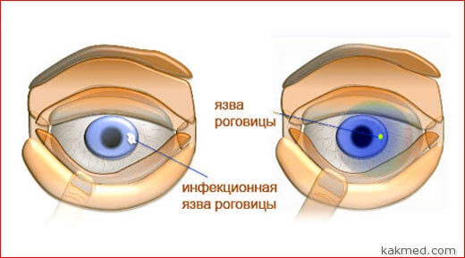
Corneal ulcer
Ulcerative keratitis or corneal ulcer is a hole in the cornea of the eye caused by a bacterial infection. Such ulcers occur after eye damage, head injuries and certain other types of damage to health.
Symptoms of ulcerative keratitis include:
- eye redness
- eye pain
- enhanced fluid secretion from the eyes
- excessive sensitivity to light
- blurred vision
- white or whitish spot on the cornea.
People suffering from eyelid ailments and those who wear contact lenses, especially if they are not very clean, are at risk for a corneal ulcer. Contaminated lenses rub the surface of the eyes, slightly damaging the upper layers of the cells. These injuries are enough to penetrate the healthy tissue of bacteria. Among the microbes that cause ulcerative keratitis are Staphylococcus aureus, E. coli, gonorrhea and pneumonia pathogens.
Therefore, contact lenses require careful handling and maintenance.

If time does not begin treatment of ulcerative keratitis, the disease will lead to loss of vision and even physical loss of the eye. In the treatment of corneal ulcers used antibiotics and antifungal drugs. That the ulcerated eye was ill less and in order to avoid complications special eye drops can be prescribed. In severe cases, a cornea transplant will be required.
Dry eye syndrome
Dry keratitis, also called dry eye syndrome, occurs in people whose body produces insufficient tears or their quality does not ensure proper wetting of the eye surface. Happens and increased evaporation of tears, associated with a reduced frequency of blinking.
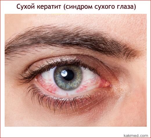
Dry eye syndrome is a symptom of a disease, such as congenital alacrimia or rheumatoid arthritis. The disease is triggered by hormonal changes and certain medications. Among such unpleasant drugs are sedatives, diuretics and antihypertensive drugs, oral contraceptives.
In chronic dry keraitis, the surface of the eye becomes irritated and inflamed, causing the eye to become red. Other dry eye symptoms include:
- burning or stinging in the eyes
- foreign body sensation
- painful sensations
- discomfort when wearing contact lenses
- blurred vision
- eye strain
- discomfort after watching TV or reading.
Only a doctor will be able to determine if any "background" disease is the cause of dry eye syndrome. If necessary, the ophthalmologist measures the amount of tears secreted by the patient’s glands, and tests for their osmolarity.
Dry keratitis is incurable, but it is possible to make life easier with non-prescription medicines, such as artificial tears (used every few hours), lubricating tear ointments (usually used during sleep), eye drops for symptomatic therapy. If the disease has gone too far, surgery is suggested.
Among the drugs, which in principle need a prescription, with dry eye syndrome is often prescribed in the form of an emulsion. It is an immunosuppressant with a superficial anti-inflammatory effect.

To protect the eye from drying out of the conjunctiva, scleral lenses or prostheses of the eye surface ecosystem are used.
Subconjunctival hemorrhage
The mucous membrane of the eye contains many blood vessels and capillaries. They can be damaged, causing blood to flow into the tight space between the conjunctiva and the white of the eye. A small amount of blood is collected under the mucous membrane. The phenomenon is known as subconjunctival hemorrhage (hypospagm). At the same time, bleeding areas give the eyes a noticeable redness.
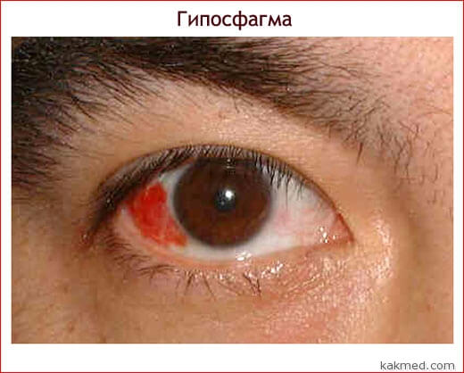
Conjunctival haemorrhage usually occurs due to minor damage or injury to the eye. Even excessive scratching of the eye with a finger can lead to hemorrhage. Those patients who have not fought or rubbed their eyes with their hands experience eye hemorrhages due to severe coughing, vomiting or a runny nose, weight lifting, or eyestrain. Subconjunctival bleeding can also occur in patients with diabetes or hypertension, as well as due to certain medications.
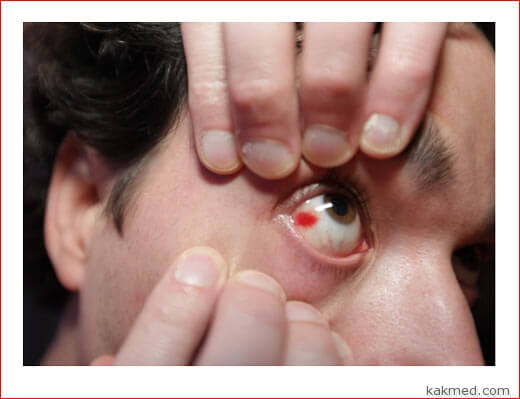
Hemorrhages under the conjunctiva occur on the surface of the organ of vision, they do not touch the cornea or the inner parts of the eye, so the vision is not impaired. Despite the frightening appearance, the bloody spots in the eyes are completely harmless, if they do not cause pain. For a few days they pass by themselves.
Other causes of eye redness
To the above reasons for the redness of the eyes in humans should be added a few more:
- inflammation of the cornea, iris or fibrous membrane of the eyeball
- glaucoma
- long stay in the bright sun
- eye contamination by dust or other particles
- swimming
- smoking and drinking
- eye irritation with any gas.
Red eye treatment
Redness of the eyes may occur from time to time or by accident. In most cases, OTC eye drops, sold in pharmacies, help. If redness does not go away after drops and is accompanied by other symptoms, see a doctor. It is possible that the cause of eye irritation was excessive "passion" for eye drops.
If, antibiotics, creams and ointments, as well as tablets can be added to the eye drops. Most illnesses causing redness of the eyes are treatable without consequences for the organs of sight.
Red eyes are a symptom of a serious disruption of the body, such as sarcoidosis, idiopathic arthritis and other dangerous ailments. The patient may not know about them.
The eye fills with blood most often in the event that a small blood vessel under the conjunctiva is damaged and there is an outpouring of blood. Most often this type of hemorrhage is affected by people with advanced age. In young people, this disease occurs most often as a result of the traumatic effect on the eye of various factors.
It occurs quite often. This type of hemorrhage occurs as a point typeso and common. The causes of the occurrence of such phenomena have been established quite a lot. The cause of the occurrence of such a disease can serve a variety of injuries, postoperative complications, as well as various local diseases.
Eye hemorrhage: causes and treatment
This disease can occur when rubbing the eyes with hands and falling into them in the process of rubbing foreign objects. Often, injury occurs through a soft corner of the pillow during sleep.. Injury of the eyeball can occur in everyday life when washing dishes with a stiff sponge, if a small piece of metal falls into the eye area. At the first stage of injury, it is difficult to determine the cause of pain arising in the eye. Ophthalmologists are often not able to make the correct diagnosis at the initial stage.
This hemorrhage often occurs together with hemorrhagic diathesis. This is determined by the color of the eye - it first appears a dark red color, after some time brightens. If the patient has problems with blood vessel sclerosis, then there is a high probability of subconjunctival hemorrhage. Foreign particles that penetrate deep into the eye, cause subconjunctival hemorrhage.
Sometimes coughing can be the cause of blood in the eye.. In addition, the cause of conjunctival hemorrhage can be excessive physical activity, a sharp jump in pressure. The redness of the eyeball also causes the use of certain coagulant preparations. It may also be caused by the use of aspirin and similar drugs. Sometimes the occurrence of hemorrhage is associated with a lack of vitamin K, as well as in violation of the mechanism of blood coagulation. Hemorrhage can have unpleasant consequences for the normal functioning of the eye, for this reason, it requires an appeal to a specialist. The eyeball with conjunctival hemorrhage does not look very nice. There is an accumulation of blood under the surface of a thin and translucent conjunctiva. In fact, the accumulation of blood is a bruise, localized in the eyeball. Hemorrhage in the eye resolves gradually independently over a period of one to three weeks. Sometimes a hemorrhage in the eyeball, together with malaise, is the result of diseases that are associated with pathologies of the blood and blood vessels.
In the treatment of subconjunctival hemorrhage, first of all, it is necessary to get rid of the underlying disease that caused the onset of hemorrhage and only after that to eliminate the hemorrhage. In order to speed up the process of resorption of blood in the eyeball drops of potassium iodide having a potassium iodide concentration of 2-3%.
This drug has a good resorption effect for blood accumulations. A very important point in the treatment of this ailment is the use of fortifying and vitamin therapy. Especially useful in the treatment is the use of ascorbic acid and vitamin P. The use of the drug askarutina allows you to replenish the body stocks of both of these vitamins, which help strengthen the vascular wall.
Do not forget to monitor your health and take seasonally. Then you can easily avoid many diseases of the blood and liver.
- In contact with 0
- Google+ 0
- OK 0
- Facebook 0