Amblyopia is a form of lowering of visual acuity, in which one eye is partially or fully not activated. Because of this, it is also called the "lazy eye" - one eye seems to be "lazy" and does not fulfill its function. As a result, the better developed eye becomes the leading one, and the second is even more inhibited.
Compared with other disorders, such as myopia, for example, “lazy eye” is a relatively rare disease and occurs in about 1–1.5% of the general population.
The priority tasks of low vision are. The projects should develop ophthalmologic facilities at the secondary and tertiary levels, which will now offer services adapted for adults and children with visual impairments, for example: the provision of appropriate equipment and materials, staff training, development of assistance programs and training. Projects can also support low-maturity regional training workshops on services. They should help develop existing services, including innovative projects aimed at raising awareness among new populations. Awareness, monitoring and evaluation. . Many visual impairments are caused by the natural evolution of vision or eye disease.
It is better to understand what amblyopia is, one can only understand its causes. Specialists distinguish the following classification of amblyopia:
Refractive amblyopia
As the name implies, refractive amblyopia arises from a violation of refraction, which leads to an inability to clearly focus on the subject. The reason may be any violation of refraction: myopia, astigmatism, hyperopia or anisometropia.
Some of them relate more specifically to older people. These symptoms may be highlighted by your ophthalmologist during your examination. However, a quick and easy test can help you detect the first signs of a disease. Look at the mosaic of your bathroom for a few minutes. If the lines are deformed, this is a sign that must be consulted without delay.
Due to a violation of the optic nerve, this pathology leads to a decrease in the field of view. The most common chronic glaucoma that develops very slowly with a progressive onset of visual discomfort. This leads to a loss of peripheral vision, and then to the central one. If in doubt, consult an ophthalmologist immediately, because without treatment, visual loss will be irreversible.
Disbiocular amblyopia
Disbinocular amblyopia occurs when there is an existing strabismus. The brain can not combine the images coming from two eyes into one, as a result of which the development of one eye stops, a "lazy eye" arises. 
Severe pain sometimes spreads to the hemisphere, accompanied by nausea and vomiting. Photomotor reflexes are canceled. The globe is hard to touch and very painful. The treatment is to administer emergency acetazolamide intravenously and refer the patient to a specialist who will inoculate miotic eye drops. Surgery is often necessary; it is peripheral iridectomy, laser or surgical.
Decreased visual acuity occurs more slowly. There is a red percussion circle. The pupil is miosis. Earth is not difficult to touch. The treatment will be applied by a specialist. The patient notices the sudden appearance of a red veil, allowing the light perception to pass.
Obscuration amblyopia, it is deprivation
Obstructive amblyopia appears due to prolonged deprivation (lack of) vision in one eye. Visual deprivation can occur due to cataracts, corneal clouding, ptosis, and other disorders that block access of light to the retina. Because of this, the eye is inactive and becomes "lazy." Even if the primary cause disorder is cured, the difference in the development of the eyes will still remain. Such amblyopia in adults takes on particularly severe forms - if untimely diagnosis and treatment, a person may be at risk of disability.
The specialist will notice intraocular bleeding, you will need to look for the cause of this bleeding, tearing the retina. Treatment will be the prerogative of the specialist. This is severe unilateral blindness. Despite rapid treatment, this blindness is very often final. Causes of obliteration of the retinal artery.
This cruel fall is always one-sided. This drop in sharpness is variable: either near or far, or both at the same time. A fixed impression, an impression of tasks, the existence of a diplopia is possible. Evolution can be done more or less quickly to complete the commitment of surgery.
Hysterical amblyopia
Hysterical amblyopia is psychogenic in nature. Occurs with prolonged hysterical amaurosis of one eye. May enter into a whole "cocktail" of psychogenic vision disorders, including color blindness, narrowing of the visual field, photophobia.
Amblyopia is often found in people suffering from diseases of the visual apparatus, inherited, as well as diseases such as Kauffman syndrome, Bench syndrome and ophthalmoplegia, as these diseases are usually accompanied by strabismus. However, the “lazy eye” itself is not inherited and does not affect the chance of its formation in descendants.
The macula must be reached to cause a decrease in visual acuity. The first signs of detachment are: - false luminous prints, for example, bright flashes - feeling of flying flies - undulating vision - sometimes amputation of the visual field. Treatment is surgical, associated with multiple processes. Insist on prophylactic treatment, which is the creation of laser coagulation, gluing around early lesions. Compression, for example, due to a benign or malignant tumor.
The test allows you to measure the visual acuity of a child from the first months of life. This test allows you to note the progress or stagnation of visual acuity and, thus, modulate the correction. The new decree provides for the measurement of visual acuity after 9 months, which suggests that this test will be a common practice among orthoptists.
Degrees of amblyopia
Many people ask: Is the "lazy eye" a reason to get a disability?
Depending on the severity of visual acuity, there are three degrees of amblyopia:
- Weak with visual acuity from 0.8 to 0.4
- Average with visual acuity from 0.3 to 0.2
- High with visual acuity from 0.1 to 0.05
- Very high from 0.04 and below
Even a high degree of amblyopia does not always mean disability - provided that the second eye sees well.
This test is to present a child with uniform gray panels, where one half is marked by a change in the circular white and black stripes. All the smaller cards are presented to the child, which allows us to mark the most beautiful network to which the child is looking.
This test is based on the fact that the infant is spontaneously attracted by a structured image resting on a neutral background. Of course, this test cannot be done without an ophthalmologic examination, but it allows detecting visual acuity below the norm depending on age.
There are also several types of this disease by fixing the eye:
- With proper fixation
- Intermittent fixation
- With stable off-center fixation
- With unstable off-center fixation
- Missing commit
Patients with correct fixation have a much more positive prognosis during treatment. Treatment of patients with improper visual fixation requires a special approach.
Hearing loss is a serious obstacle: one of three subjects after 60 years is affected. Treatment, prosthetics or re-education should be taken into account. Prebiotic or hearing loss is called hearing loss. This decrease in hearing acuity is peripheral, that is, it affects the organ of hearing, and not the region of the brain.
It is selective, that is, it does not affect all sounds equally. This hearing loss is often unknown, as it is gradually being established, allowing the patient some addiction. But this hearing disorder must be put at an early stage, because language is a tool for communication, and this can jeopardize all relationships with others, hence the tendency to withdraw into oneself.
Diagnosis and treatment in children
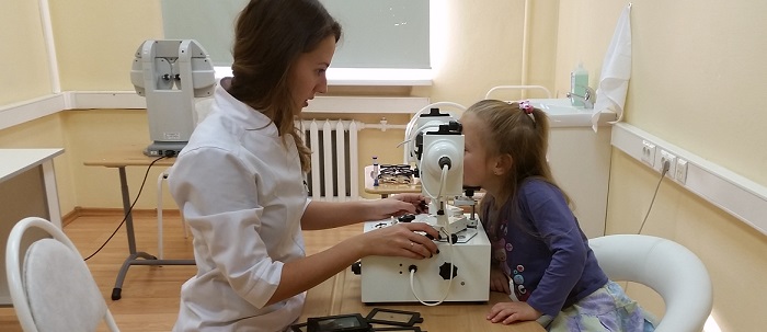
Amblyopia in children is most often found during a medical examination at school or kindergarten. If the "lazy eye" is detected in the early stages of development, the treatment will be relatively simple and quick. If you suspect this disease, the child undergoes a more thorough examination by an ophthalmologist, which includes: visometry, refractometry, ophthalmometry, biomicroscopy and other examinations. It is also possible additional examination by a neurologist.
A reversed ear can become emotional, even intellectual. Two tests allow you to make a diagnosis. Tonal audiometry, which allows you to find out the area of the cochlea that is most affected. As a rule, the one that recognizes loud sounds.
Drugs: Ergotamine derivatives act on the neuron. Prosthetics: this should no longer be synonymous with negative aging, but, on the contrary, proof of the desire to integrate and not give up. Rehabilitation: this may include the study of lip reading and auditory retraining.
The risk factors for the formation of a "lazy eye" for the most part are not inherited, most often it is:
- Prematurity, especially grade 3-4
- Mental retardation
- Strabismus
- Kauffman syndrome
- Benche's syndrome
- Early childhood retinopathy
If there is a history of any of these diseases - you should regularly examine the child with an ophthalmologist.
Gradual decrease in visual acuity
Any hearing loss after 60 years should be taken very seriously by the attending physician before the disability becomes too serious. All of the following pathologies are most often distinguished by an ophthalmologist during normal visits or when certain chronic conditions are observed. Their treatment is often associated with their causative pathology.
It is the attachment of the eye, leading to partial or full opacity of the lens. This clouding may be congenital, traumatic, toxic, diabetic or age-related. Age-related cataracts are the most common causes.
Amblyopia in children responds well to treatment, especially in mild form. The treatment of this disease is called "pleoptic treatment" and is aimed at restoring normal binocular vision.
The basis of treatment is occlusion - this method has been used for 200 years and has proven its undeniable effectiveness. It consists in closing one eye with the help of an occluder - a special bandage or blind cap for glasses. Occlusion can be:
The only treatment is the use of surgery. It consists of removing the opaque lens and replacing it with an implant. The technique of this intervention is currently very well controlled. The results are very satisfactory, and this intervention can be performed on an outpatient basis without hospitalization. There are no age restrictions for its use.
This is an abnormality with increasing eye pressure, most often developing in a cunning and painless way. Untreated, it amputates the progressive loss of the visual field after slow destruction of the optic nerve, which can lead to complete loss of vision.
- Direct (closes better seeing the eye)
- Reverse (closes the worse seeing eye)
- Alternating (both eyes close in turn)
According to the duration of the closing of the eye, a constant is distinguished (the eye is closed all the time), partial (it is allowed to open the eye for a while) and minimum (the eye is closed rarely, during some specific types of activity).
This is a condition that most often affects people over 40 years old. This is very common, and the frequency increases with age. Many drugs are contraindicated. Treatment is most often medical and is aimed at normalizing intraocular pressure. In some cases, surgical treatment may be required.
This is a loss of central vision due to deterioration of the macula. This is the first cause of blindness for people over 50 years old. In France, we are talking about a million people. Early detection is important for the implementation of laser treatment, which can be effective in forms diagnosed early enough. Recently, a new drug has been approved for the treatment of certain forms. It is reimbursed by social security if special conditions are met. It is used for intravenous administration in combination with laser photobased.

It is necessary to ensure that the child does not remove the occluder - a violation of the regime can put the months of treatment down the drain. Particularly often, parents of high schoolchildren face this problem; they are embarrassed to wear an occluder, not realizing that they put themselves at risk of becoming disabled.
Progressive retinal changes resulting in loss of vision. It can be familial, hereditary, and bilateral, or after diabetes, high blood pressure, or drug poisoning. There is another form, retinitis pigmentosa, characterized by degeneration of the rods.
The defect is in how the optical system forms the images, as a result of which the light rays from a point object cannot form an ideal point of the image. The peripheral zones of the most common progressive slow lenses show unwanted aberrations of this type.
The duration of occlusion may be different, depending on the severity of the disease, but as a rule it is from 1 to 5 years. This therapy requires high responsibility, both on the part of the doctor and on the part of the parents, because improperly organized or disturbed occlusion can have the following side effects:
- Decreased visual acuity of a healthy eye
- Strabismus development
- Diplopia (double vision)
- Dermatozy and other cosmetic defects
Optical vision correction may also be applied. Depending on the type of amblyopia, both glasses and contact lenses are used. With the help of optical correction can be performed so-called penalization, when the patient artificially develops anisometropia. The advantage of this method is that a healthy eye is involved in the process of vision and does not lose sharpness.
The ability of the eye to clearly see objects at close range due to changes in the crystalline curvature. The ability of the eye to distinguish the details of an object. The minimum resolution or resolution angle expresses the smallest distance between two lines, which is perceived as two separate objects.
Optical power required for careful viewing, in addition to that required for viewing from afar. Visual fatigue is characterized by acute or acute eye irritation, blurred vision and headache, most often at the end of the day.
As an additional therapy, eye gymnastics, various medications and video-computer treatment methods can be prescribed.
In the case of the psychogenic form of the disease, an appointment to a psychotherapist or psychiatrist is also possible.
With timely treatment, amblyopia in children (up to 7 years) has a very favorable prognosis. As a rule, it is possible not only to preserve, but almost completely normalize visual acuity. After 11-12 years, treatment will be much more difficult and long, and the result will be insignificant.
The perpendicular distance between the center of the pupil and the lower part of the selected frame. Amblyopia is also called the “lazy eye,” a decrease in visual acuity diagnosed in childhood. It contains all visual impairments that prevent the formation of a “clear image on the retina,” with the exception of age-related presbyopia. Myopia, hyperopia and astigmatism are all forms of ametropia.
This is a condition in which two eyes have different refractions. Anomaly caused, in most cases, by the uneven curvature of the cornea, which causes, therefore, a distorted view of the objects. Bars are photoreceptors in the retina that provide night vision, respond to light, but cannot distinguish colors.
Diagnosis and treatment of amblyopia in adults
Amblyopia in adults is often much more difficult, mainly due to the neglect of the disease - people often go to the doctor only when they notice that they have severe visual impairment. More often than in children, the hysterical form of this disease occurs.
Traditional forms of treatment, such as occlusion, have almost no effect on adults. Therefore, doctors often use various hardware and medical treatment methods.
Corrective lenses designed to correct presbyopia. They include two main areas. The bottom of the lens provides closer attention; the top is a vision from afar. The intermediate zone is killed. The crystal is opaque, that is, a natural lens inside the eye that resolves with the extraction of the lens and its replacement, most often with an intraocular lens. A cataract can also be hereditary or the result of an injury.
The condition of the person who is blind, i.e. devoid of any visual perception. The conjugated photosensitive neurons found in the retina are used for central vision and color perception. Transparent front of a spherical or spherical eye globe. In combination with crystalline, the cornea plays an important role in focusing images on the retina.
The prognosis for treating a “lazy eye” in adulthood is very unfavorable. Even a very long treatment can give a poorly sensible result, the probability of a relapse is high. The situation can be exacerbated by strongly pronounced squint.
Therefore, do not forget to be regularly examined by an ophthalmologist and brought to the inspection of your children, because timely diagnosis of the disease is the key to successful treatment!
Rapidly developing reduction in visual acuity in young people is most often the result of retrobulbar neuropathy of the optic nerve, or retrobulbar neuritis, which occurs more often on the one hand, sometimes on both sides, simultaneously or sequentially.
Clinical manifestations. The patient first notes the appearance of fog, a veil in front of the eye, sometimes pain in the depth of the orbit. Decrease in visual acuity while usually increasing over several hours or several days, can reach 0.1-0.2; complete blindness rarely occurs. The pupil on the side of the pathological process can be expanded, its reaction to light is reduced. The cause of this form of pathology is often demyelination of the optic nerve fibers. Due to the retro-bulbar localization of the process, there may be no abnormalities in the fundus, but often with ophthalmoscopy there are signs of papillitis: hyperemia and elevation (elevation) of the optic nerve head into the vitreous; however, there is no significant hyperemia of the retinal veins. The latter circumstance helps in ophthalmoscopy to distinguish papillitis from the stagnant optic nerve head. The main criterion for their differentiation is simple: with retrobulbar neuritis, in particular with papillitis caused by it, a decrease in visual acuity occurs acutely or subacutely and is very pronounced, whereas with a stagnant disc, visual acuity does not change for a long time (weeks, months) or changes remain insignificant.
In case of retrobulbar neuritis, there may be some soreness during the movements of the eyeballs and pressure on them. Characterized by predominant loss of central vision, while peripheral vision may in that
or to a certain extent remain safe. One of the main causes of retro-bulbar neuritis is autoimmune inflammation, accompanied by demyelination of the optic nerve fibers.
Restoration of visual acuity usually occurs within a few weeks after its decrease, appears gradually, and as a result, vision is restored, usually reaching its initial level. In rare cases, a small central scotoma remains in the visual field of the affected eye. Later, some blanching (atrophy) of the optic nerve head, especially significant on its temporal side, can be detected. Manifestations of retrobulbar neuritis may recur.
In persons who have undergone retrobulbar neuritis, further, within 10–15 years, symptoms of demyelination develop in 20–40% of cases in other parts of the CNS, which makes it possible to diagnose a common demyelinating disease - multiple sclerosis, which is the most common cause of retrobulbar neuritis.
Bilateral optic neuritis can precede the development of the clinical picture of transverse myelitis by several days or weeks. The combination of retrobulbar neuritis on both sides and acute transverse myelitis is known as acute opticomyelitis, or Devic's disease.
Other, in addition to multiple sclerosis, the causes of unilateral retrobulbar neuritis can be acute disseminated encephalomyelitis, posterior uveitis, viral infections, sarcoidosis.
Treatment. In case of retrobulbar neuritis, treatment with corticosteroids is indicated, which can be administered retrobulbarly. Pulsed therapy with methylprednisolone is also effective (1000 mg in 200 ml of 5% glucose solution intravenously drip 1 time a day for 3 days, followed by a rapid dose reduction and switching to the intake of prednisozolone orally at 1 mg / kg / day, followed by a gradual decrease in the daily dose by 5 mg per day). This treatment speeds up the restoration of vision, but the likelihood of developing a further developed clinical picture of multiple sclerosis is retained.
Transient monocular blindness - bouts of rapidly developing uniform decrease (darkening, extinction) of vision in one eye.
Etiology and clinical manifestations. Often within 10–15 s, blindness develops, usually lasting from a few seconds to a few hours, sometimes recurring in nature and not accompanied by pain. Lshaigo818 Guides (Greek Ashaigo818 - darkening, Guides - fleeting) is usually the result of transient retinal ischemia, most often occurring against the background of stenosis of the internal carotid artery. In such cases, monocular blindness or a pronounced short-term reduction of vision in one eye is a symptom of ophthalmogram plexus syndrome, in which it is accompanied by transient central hemiparesis on the opposite side. This syndrome may be a precursor of ischemic stroke.
With transient monocular blindness or reduction of vision in one eye, caused by stenosis of the internal carotid artery, noise is possible during auscultation of this vessel. Diagnosis confirmation can be provided by USDG and duplex scanning, as well as carotid angiography. Tactics of treatment is usually determined based on the data obtained from the application of these diagnostic methods.
Monocular blindness is sometimes the result of microemboli of retinal arteries (arterio-arterial or cardioarterial microemboli), as well as stenosis of the central retinal artery, anterior ischemic neuropathy of the optic nerve caused by giant cell arteritis or atherosclerotic lesion of the ophthalmic artery or its branches.
Anterior ischemic optic neuropathy is characterized by acute painless loss of vision in one eye; in severe cases, the resulting blindness may be persistent; in the fundus there may be swelling and blanching of the optic nerve head, peripapillary foci of hemorrhage, the retina of the eye remains unchanged.
Occlusion of the central retinal artery may manifest sudden blindness in one eye. Its retina is pale in the ischemic area, at the location of the yellow spot it can be so thin that sometimes the choroid membrane appears through it - a red spot is detected, known as a symptom of a cherry stone.
Transient monocular blindness may be the aura of the retinal form of migraine, which is characterized by dysgemic manifestations in the system of retinal arteries. In such cases, a decrease in visual acuity in one eye is soon accompanied by a characteristic headache, usually on the same side as a hemicranium, especially intense in the frontal-orbital region. Sometimes patients with a more common ophthalmic form of migraine also complain of a loss of vision in one eye during a migraine attack, however, in such cases, clarification of the history allows to establish that the aura actually manifested itself in the form of a homonymous hemianopsia, usually explained by vasospasm in the posterior cerebral artery pool . By the way, in case of ophthalmic migraine, photopsies and shimmering scotomas are often observed in the homonymous halves of both eyes, and hemicrania attacks usually occur on the opposite side. Manifestations of the visual aura (photopsies, scotomas, narrowing of the visual fields, temporary decrease in visual acuity, sometimes even blindness), spreading across the entire field of view, can occur on both sides. This is characteristic of attacks of ophthalmic migraine, during which vascular reactions occur in the basilar artery or in both posterior cerebral arteries.
Toxic metabolic neuropathy of the optic nerves is usually manifested by simultaneous deterioration of vision on both sides. At the same time, central or centrocecal (central, merging with a blind spot) scotomas form within a few days or weeks.
Etiology and clinical manifestations. Toxic metabolic neuropathy in such cases can be caused by intoxication with levomycetin, streptomycin, sulfonamides, isoniazid (tubaside), digitalis, digoxin, arsenic, lead, thallium, teturamo (antabuse). In cases of poisoning with methyl alcohol (methanol), vision disorders appear on the background of intoxication in the form of acute large symmetrical central cattle or complete barely sweats, as well as other signs of neurological and somatic pathology, acidosis (see Chapter 46).
Metabolic neuropathy of the optic nerves can be due to a deficiency of folic acid, vitamins B] and B] 2, usually associated with inadequate nutrition, malabsorption, starvation, and alcoholism.
Degenerative changes in the retina of the eyes and optic nerves are a possible consequence of a number of hereditary diseases, in particular Leber's disease (Leber's optic neuropathy) inherited in the recessive, linked to the X chromosome type and therefore occurring only in men. In Leber's disease, amaurosis can be congenital, or visual impairment debuts later, before the age of 30. In such cases, the visual field narrows in parallel and its sharpness increases up to blindness. Ophthalmoscopic picture may vary widely. In particular, signs of primary atrophy of the optic nerve discs, narrowing of the arterioles of the retina, pigmentation of the fundus of the eye, the appearance of granularity in the fundus of the yellow spot, diffuse white lesions, signs of chorioretinal atrophy can be detected. On electroretinograms marked pronounced changes characteristic of tapetoretinal degeneration.
Severe visual impairment, including blindness due to damage to the retina and optic nerve, is one of the most important signs of a group of lysosomal diseases belonging to gangliosidoses and inherited in an autosomal recessive manner, which until recently were known as amaurotic idiocy (Tay-Sachs disease and others).
Finally, senile macular degeneration and various forms of retinitis pigmentosa, retinal degeneration and detachment, cataracts and other diseases and traumatic injuries of the refracting medium of the eye lead to reduced vision, sometimes blindness.
From a decrease in vision and blindness (amaurosis) from early childhood, amblyopia is significantly different - a decrease in vision due to the functional features of some structures related to the visual analyzer. Options for amblyopia: a) anisometropic amblyopia - character
it is tested by poorly corrected decrease in visual acuity of the eye with more pronounced ametropia (refractive error); b) refractive amblyopia due to refractive error (mainly during high degree hypermetropia and astigmatism), which is not amenable to optical correction; c) amblyopia due to anopia is the result of functional inactivity of the eye, for example, with pronounced monolateral strabismus and constant inhibition in connection with this function of the central vision of the mowing eye.
- In contact with 0
- Google+ 0
- OK 0
- Facebook 0